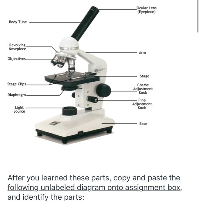
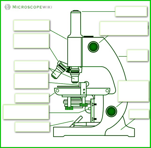
41 diagram of a microscope with labels
PDF Label parts of the Microscope: Answers Label parts of the Microscope: Answers Coarse Focus Fine Focus Eyepiece Arm Rack Stop Stage Clip The Parts of a Microscope (Labeled) Printable Printable ... The Parts of a Microscope (Labeled) Printable. This diagram labels and explains the function of each part of a microscope. Use this printable as a handout or transparency to help prepare students for working with laboratory equipment.
Microscope, Microscope Parts, Labeled Diagram, and Functions The slide is the bottom layer. The liquid sample comes next. To minimise evaporation and protect the microscope lens from sample exposure, a small square of clear glass or plastic (a coverslip) is placed on top of the liquid. 1. Collect a clean microscope slide and a coverslip (a thin piece of plastic covering).

Diagram of a microscope with labels
en.wikipedia.org › wiki › Electron_microscopeElectron microscope - Wikipedia An electron microscope is a microscope that uses a beam of accelerated electrons as a source of illumination. As the wavelength of an electron can be up to 100,000 times shorter than that of visible light photons, electron microscopes have a higher resolving power than light microscopes and can reveal the structure of smaller objects. Microscope Labeling - The Biology Corner Students label the parts of the microscope in this photo of a basic laboratory light microscope. Can be used for practice or as a quiz. Label Microscope Diagram - EnchantedLearning.com Label Microscope Diagram Using the terms listed below, label the microscope diagram. Inventions and Inventors arm - this attaches the eyepiece and body tube to the base. base - this supports the microscope. body tube - the tube that supports the eyepiece. coarse focus adjustment - a knob that makes large adjustments to the focus.
Diagram of a microscope with labels. Microscope Poster - Diagram with Labels | Teach Starter A poster containing a diagram with labels showing the key parts of a microscope. Use this educational classroom poster in your science lessons to highlight the key parts of a microscope. A lot of equipment is used in science experiments and it is important to know the names of and understand each part of the equipment and how it works. bodycoach-online.de › morphology-tree-diagram-exercisesMorphology tree diagram exercises - bodycoach-online.de 1 day ago · Center for the Advancement of Faculty Excellence (CAFE)Tree diagrams. usssa registration fee 2020 Home; nail salon greensboro, nc open sunday ContactA tree diagram, or tree, is a two-dimensional diagram used in generative grammar as a convenient Spring 2012, April 5 Missing morphology Variability in acquisition Morphology and functional ... PDF Parts of a Microscope Printables - Homeschool Creations and 40x. The eyepiece on a microscope magnifies at 10x, so when used together, the 4x lens magnifies an item 40x, the 10x magnifies 100x, and the 40x magnifies 400x. (note: for typical student microscope -other microscopes will vary) •Which part of the microscope rotates so another person can look through the eyepiece Labelled Diagram Of A Plant Cell Under A Microscope ... Animal Cell Diagram Electron Microscope. 11 is a labelled diagram of a leaf palisade mesophyll cell as seen With a high quality light microscope. But at the same time. Under the microscope Priya observes a cell that has a cell wall and a distinct nucleus. Its a thin slice.
Draw a labelled diagram of an image formed by a compound ... Click here👆to get an answer to your question ️ Draw a labelled diagram of an image formed by a compound microscope, with the image at least distance of distinct vision. Write any one expression for its magnifying power. Parts of a Compound Microscope - Labeled (with diagrams ... It is used to carry the microscope and at the same time connect the base of the microscope to the head. (1, 2, 3, and 4) Image 3: A compound microscope with a corresponding label of the different parts. quizlet.com › 568033559 › botany-exam-1-chs-1/2/3-4Botany Exam 1 Chs. 1, 2, 3, 4, 5, 6, 7, 12, and 16 Quizzes ... Mitosis, or the division of a mother cell's nucleus into two identical daughter nuclei, is typically divided into four phases. Match each of the labels to identify what events take place during each phase of mitosis. 1. Prophase 2. Metaphase 3. Anaphase 4. Telophase A) Chromosomes are aligned at the equator of the cell and the spindle is fully ... Parts of a Simple Microscope - Labeled (with diagrams ... A simple microscope is a very first type of microscope ever created. It consists of simple parts and performs simple functions. Although there are now many advanced microscope types, some applications may still demand the use of a simple microscope. In this article, we are going to discuss the parts and functions of a simple microscope.
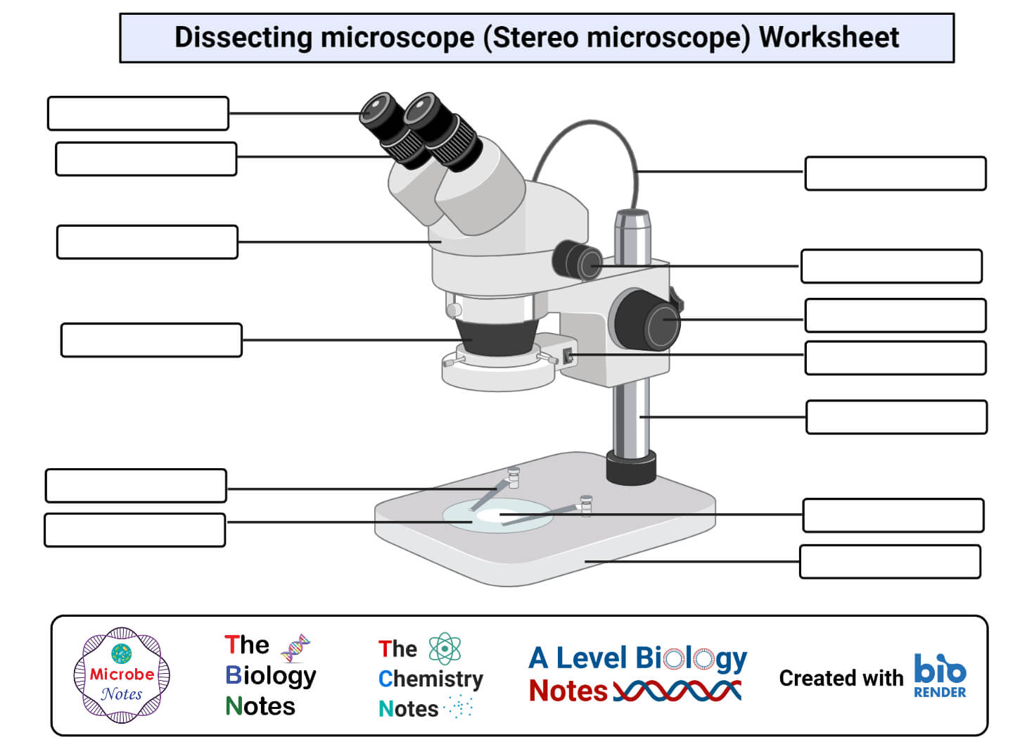
rsscience.com › stereo-microscopeParts of Stereo Microscope (Dissecting microscope) – labeled ... Labeled part diagram of a stereo microscope Major structural parts of a stereo microscope. There are three major structural parts of a stereo microscope. The viewing Head includes the upper part of the microscope, which houses the most critical optical components, including the eyepiece, objective lens, and light source of the microscope. Diagram of a Compound Microscope - Biology Discussion Information recorded on adhesive label is stuck to the base of the microscope for future reference. (ii) Use: Having calibrated the eyepiece scale for all the objective lenses on the microscope, one can use it to measure the dimensions of cellular and sub-cellular structures, e.g., bacterial cells, fungal spores onion epidermal cells etc. Microscope Drawing And Label at PaintingValley.com ... Tags: microscope, label All rights to paintings and other images found on PaintingValley.com are owned by their respective owners (authors, artists), and the Administration of the website doesn't bear responsibility for their use. Label the Light Microscope - Labelled diagram Label the Light Microscope. Share Share by Nquinn805. Like. Edit Content. Embed. More. Leaderboard. Show more Show less . This leaderboard is currently private. Click Share to make it public. This leaderboard has been disabled by the resource owner. This leaderboard is disabled as your options are different to the resource owner. ...
Parts of a microscope with functions and labeled diagram
Labeling the Parts of the Microscope | Microscope World ... Labeling the Parts of the Microscope. This activity has been designed for use in homes and schools. Each microscope layout (both blank and the version with answers) are available as PDF downloads. You can view a more in-depth review of each part of the microscope here. Download the Label the Parts of the Microscope PDF printable version here.

Conceptual zoology aspirants - Microscope diagram | Facebook
Compound Microscope Parts - Labeled Diagram and their ... Labeled diagram of a compound microscope Major structural parts of a compound microscope Optical components of a compound microscope Eyepiece Eyepiece tube Objective lenses Nosepiece Specimen stage Coarse and fine focus knobs Rack stop Illuminator Condenser Abbe condenser Iris Diaphragm Condenser Focus Knob Summary An overview of microscopes

GCSE Optical microscope labelling Diagram | Quizlet
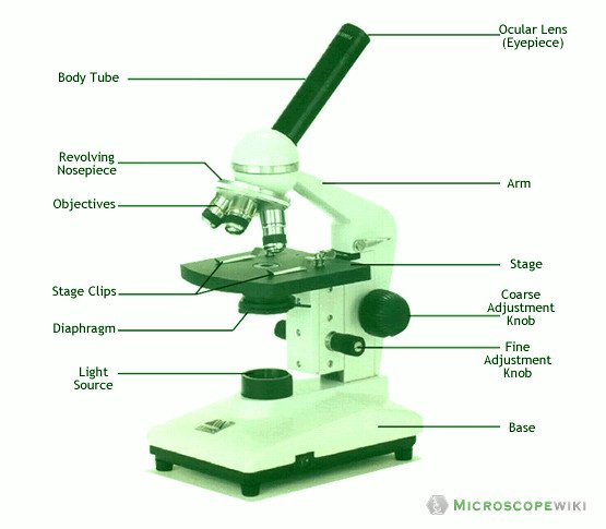
Parts of the Microscope with Labeling (also Free Printouts ... Image 2: The body tube part of a microscope is where the ray of light is bent to allow the object being viewed to enlarge by the scope. Picture Source: slideplayer.com 3. Turret/Nose piece. It is the revolving part of the microscope. It allows the use of different types of objective lenses by simply rotating the top part of the turret.

Microscope Diagram Labeled, Unlabeled and Blank | Parts of a ...
› books › NBK26880Looking at the Structure of Cells in the Microscope ... A light microscope. (A) Diagram showing the light path in a compound microscope. Light is focused on the specimen by lenses in the condensor. A combination of objective lenses and eyepiece lenses are arranged to focus an image of the illuminated specimen
Solved diagram shows a typical light microscope with its ...
pages.zeiss.com › rs › 896-XMS-794Principles of Fluorescence and Fluorescence Microscopy The Fluorescence Microscope The main requirement of a fluorescence microscope is to illuminate a specimen with light of an excitatory wavelength whilst simultaneously collecting and separating the compara-tively weaker light emitted by the sample. In the example of Stokes’ observation, these tasks are performed by the

File:Microscope diagram.png - Wikimedia Commons
Microscope Parts and Functions With Labeled Diagram and ... The specimen is placed on the glass and a cover slip is placed over the specimen. This allows the slide to be easily inserted or removed from the microscope. It also allows the specimen to be labeled, transported, and stored without damage. Stage: The flat platform where the slide is placed.

microscope diagram - Google Search | Diagram, Microscope ...
A Study of the Microscope and its Functions With a Labeled ... These labeled microscope diagrams and the functions of its various parts, attempt to simplify the microscope for you. However, as the saying goes, 'practice makes perfect', here is a blank compound microscope diagram and blank electron microscope diagram to label. Download the diagrams and practice labeling the different parts of these fascinating instruments.
How to Use a Microscope
PDF Parts of the Light Microscope - Science Spot Parts of the Light Microscope T. Trimpe 2003 A. EYEPIECE Contains the OCULAR lens J. COARSE ADJUSTMENT KNOB Moves the stage up and down for FOCUSING I. FINE ADJUSTMENT KNOB Moves the stage slightly to SHARPEN the image G. BASE Supports the MICROSCOPE D. STAGE CLIPS HOLD the slide in place C. OBJECTIVE LENSES

Microscope Diagram Labeled, Unlabeled and Blank | Parts of a ...
Microscope labeled diagram - SlideShare Microscope labeled diagram 1. The Microscope Image courtesy of: Microscopehelp.com Basic rules to using the microscope 1. You should always carry a microscope with two hands, one on the arm and the other under the base. 2. You should always start on the lowest power objective lens and should always leave the microscope on the low power lens when you finish using it. 3.

Label the Microscope Diagram | Download Scientific Diagram
Compound Microscope Parts, Functions, and Labeled Diagram ... Compound Microscope Definitions for Labels. Eyepiece (ocular lens) with or without Pointer: The part that is looked through at the top of the compound microscope. Eyepieces typically have a magnification between 5x & 30x. Monocular or Binocular Head: Structural support that holds & connects the eyepieces to the objective lenses.

Compound Microscope Parts
Labeled Diagram Of A Stereo Microscope | Products ... MX63 Semiconductor Inspection Microscope Olympus MX63 semiconductor industrial inspection microscope systems offer easy, accurate, and comfortable observations of micro-structures on wafers up to 300 mm. The microscopes meet international standards including SEMI S2/S8, CE, and UL. Advanced imaging technology delivers clear micrographs, and an ergonomic design helps minimize user fatigue.

Microscope Diagram Labeled, Unlabeled and Blank | Parts of a ...
Microscope Labeling Diagram - Quizlet Start studying Microscope Labeling. Learn vocabulary, terms, and more with flashcards, games, and other study tools.

Parts of Stereo Microscope (Dissecting microscope) – labeled ...
› confocal-microscopes › lsm-980LSM 980 with Airyscan 2 – Confocal Microscope with Multiplex ... LSM 980 can image multiple labels simultaneously, covering a wide emission range up to 900 nm. These Cos-7 cells were labelled with 4 different fluorophores, two of which have their emission peak in the near infrared range (NIR), Alexa 700 and Alexa 750.

Microscope Parts and Functions
Label the microscope - Science Learning Hub Drag and drop the text labels onto the microscope diagram. All microscopes share features in common. In this interactive, you can label the different parts of a microscope. Use this with the Microscope parts activity to help students identify and label the main parts of a microscope and then describe their functions.
16 Parts of a Compound Microscope: Diagrams and Video ...
Parts of Microscope | Function | Labeled Diagram ... To examine these small objects with high magnification microscope parts are made with special components with high accuracy. Due to that, accurate examination and results are possible to achieve. Microscope parts labeled diagram gives us all the information about its parts and their position in the microscope. Microscope Parts Labeled Diagram
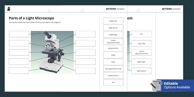
Parts of a Light Microscope Cut and Stick Worksheet - Twinkl
Parts of a microscope with functions and labeled diagram Parts of a microscope with functions and labeled diagram February 9, 2022 December 24, 2021 by Faith Mokobi Having been constructed in the 16th Century, Microscopes have revolutionalized science with their ability to magnify small objects such as microbial cells, producing images with definitive structures that are identifiable and characterizable.

Microscope, Microscope Parts, Labeled Diagram, and Functions
Simple Microscope - Parts, Functions, Diagram and Labelling Simple Microscope - Parts, Functions, Diagram and Labelling By Editorial Team March 7, 2022 A microscope is one of the commonly used equipment in a laboratory setting.
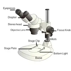
Dissecting Stereo Microscope Parts and Functions
Labelled Diagram of Compound Microscope The below mentioned article provides a labelled diagram of compound microscope. Part # 1. The Stand: The stand is made up of a heavy foot which carries a curved inclinable limb or arm bearing the body tube. The foot is generally horse shoe-shaped structure (Fig. 2) which rests on table top or any other surface on which the microscope in kept.

Draw a well labelled diagram of a microscope. - Brainly.in
Label Microscope Diagram - EnchantedLearning.com Label Microscope Diagram Using the terms listed below, label the microscope diagram. Inventions and Inventors arm - this attaches the eyepiece and body tube to the base. base - this supports the microscope. body tube - the tube that supports the eyepiece. coarse focus adjustment - a knob that makes large adjustments to the focus.
Simple Microscope - Diagram (Parts labelled), Principle ...
Microscope Labeling - The Biology Corner Students label the parts of the microscope in this photo of a basic laboratory light microscope. Can be used for practice or as a quiz.

This is a common compound microscope Label its parts class 11 ...
en.wikipedia.org › wiki › Electron_microscopeElectron microscope - Wikipedia An electron microscope is a microscope that uses a beam of accelerated electrons as a source of illumination. As the wavelength of an electron can be up to 100,000 times shorter than that of visible light photons, electron microscopes have a higher resolving power than light microscopes and can reveal the structure of smaller objects.

2.1 " Compound Microscope" | Download Scientific Diagram
Microscope Types (with labeled diagrams) and Functions

Simple Microscope - Diagram (Parts labelled), Principle ...

Compound Microscope Parts – Labeled Diagram and their ...
1.5: Microscopy - Biology LibreTexts

Carl Zeiss Microscopy Optical microscope Worksheet Diagram ...
Parts of a microscope with functions and labeled diagram

Simple Microscope Definition, Magnification, Parts And Uses

The Microscope
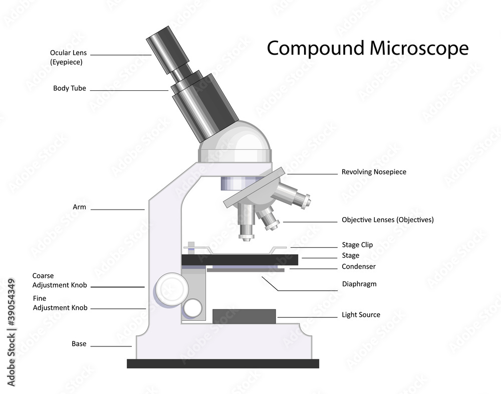
Compound Microscope with labels Stock Vector | Adobe Stock

Compound Microscope Parts, Function, & Diagram | What is a ...

Diagram Of A Microscope | Diagram, Microscope, Science ...
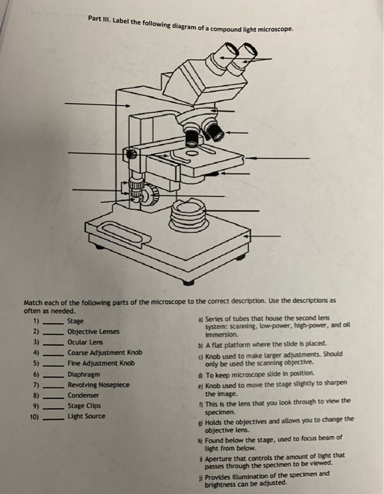
Solved Part III. Label the following diagram of a compound ...

Light Microscope vs Electron Microscope - Accelerating Microscopy
Modified Science Diagram; Label Parts of a Microscope; Special Education
Label a microscope - Teaching resources

Compound Microscope: Definition, Diagram, Parts, Uses ...

Parts of Microscope | Function | Labeled Diagram | slidingmotion

Draw a neat labelled diagram of a compound microscope class ...

Parts of the Microscope with Labeling (also Free Printouts ...
0 Response to "41 diagram of a microscope with labels"
Post a Comment