44 gram negative bacteria diagram
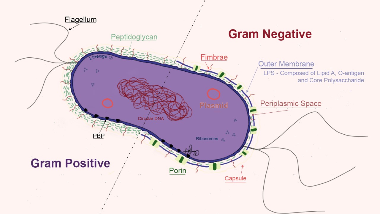
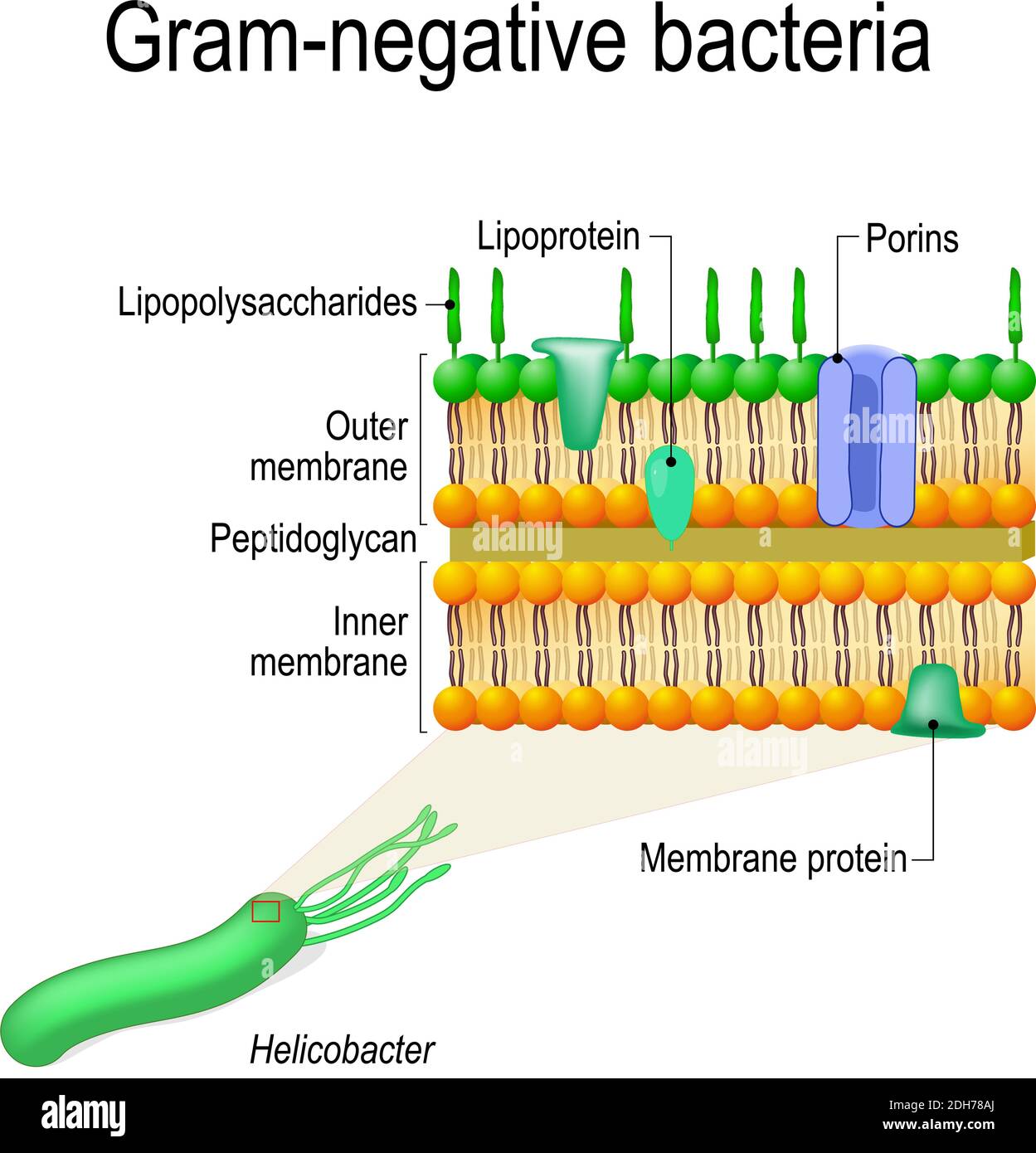
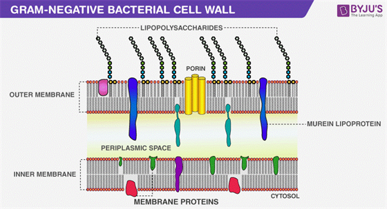
Gram-negative bacteria cell wall. The Cell wall of the Gram-Negative Bacteria is very complex as compared to that of Gram-Positive Bacteria. Combined with the major role of the outer membrane of the cell, with a layer of peptidoglycan, its functional properties are complex, and here is a description of the cell wall and its functional parts. The types of bacteria and their labeled diagrams are shown below. The sizes of bacteria cells that can infect humans beings range from 0.1 to 10 micrometers. Some larger types of bacteria such as the rickettsias, mycoplasmas, and chlamydias have similar sizes as the largest types of viruses, the poxviruses.
Klebsiella, Enterobacter, and Serratia are closely related gram-negative bacteria Overview of Gram-Negative Bacteria Gram-negative bacteria are classified by the color they turn after a chemical process called Gram staining is used on them. Gram-negative bacteria stain red when this process is used.
Gram negative bacteria diagram
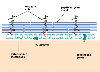
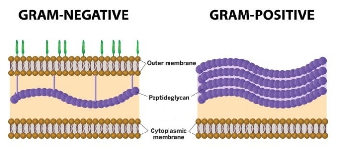
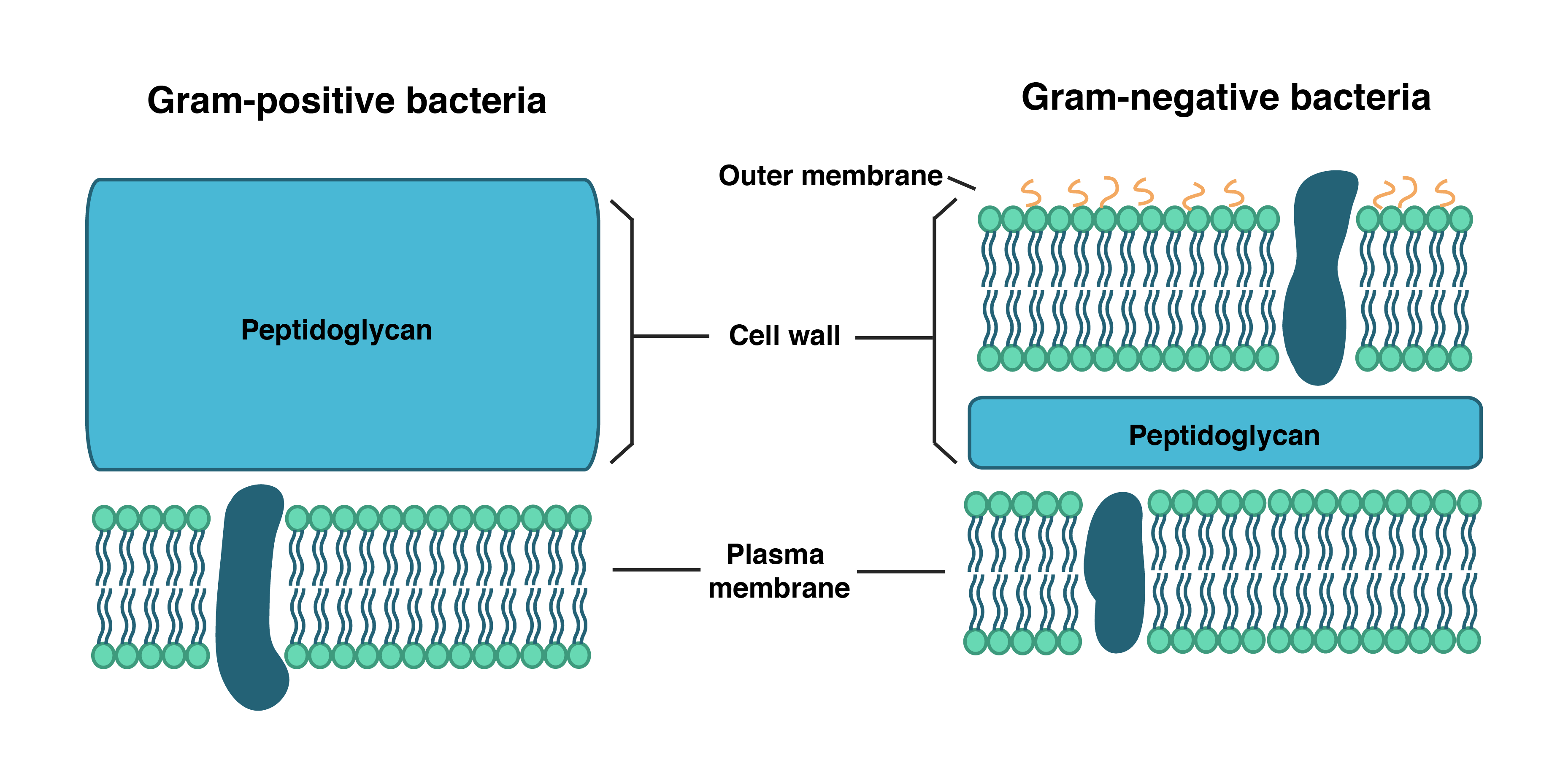
Gram-negative bacteria have a smaller amount of this rigid structure than do gram-positive bacteria. Periplasmic space. an open area between the cell wall and cell membrane in the cell envelopes of bacteria. gram negative bacteria have a more extensive space than do a gram positive bacteria. Cell membrane. Bacteria can be classified based on shape, mode of nutrition, respiration, the composition of the cell wall, etc. Based on these criteria, bacteria can be classified as bacillus, coccus or vibrio, etc., autotrophic or heterotrophic, aerobic or anaerobic, Gram + or Gram -. 8. According to Peberdy (1980) the only compound present in the cell walls of both Gram-negative and Gram-positive bacteria is 'peptidoglycan'. The cell walls of Gram-positive bacteria contain up to 95% peptidoglycan and up to 10% teichoic acids. 9. Cytoplasmic membrane is a thin (5-10 nm) layer lining the inner surface of the cell wall.
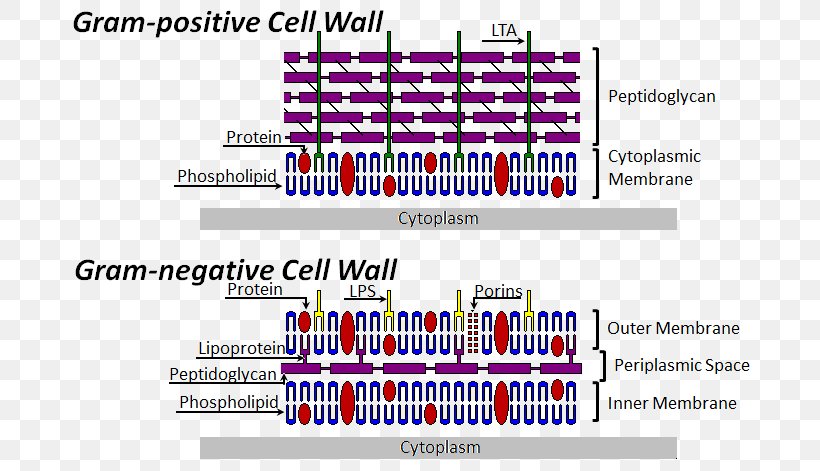
Gram negative bacteria diagram. The diagram above illustrates the differences in the structure of Gram positive and Gram negative bacteria. The two key features that lead to the differing visualization properties of Gram positive and Gram negative species are the thickness of the peptidoglycan layer and presence or absence of the outer lipid membrane. Gram-positive bacteria remain dark-violet or purple coloured, but Gram-negative bacteria become red (pink) (Fig. 2.10). This grouping does not depend on the shape of bacteria, e.g., the bacillus-shaped bacteria like Bacillus subtilis is Gram-positive, while the other rod-shaped bacteria like Coxiella burnetii is Gram-negative. Flow Chart Of The Study Design Each Patient Having A First Positive Scientific Diagram. Flowchart For Identification Of Anaerobic Gram Positive Bacilli 1 Scientific Diagram. Gram Negative Bacteria Positive Microbiology Flowchart Others Angle Text Infection Png Pngwing. Starting With Figure 17 5 And Using From Case Study T Chegg. Compare and contrast the cell walls of typical Gram-positive and Gram-negative bacteria. 3. Relate bacterial cell wall structure to the Gram-staining reaction. 37 . 38 Bacterial Cell Wall • Peptidoglycan (murein) -rigid structure that lies just outside the cell plasma membrane
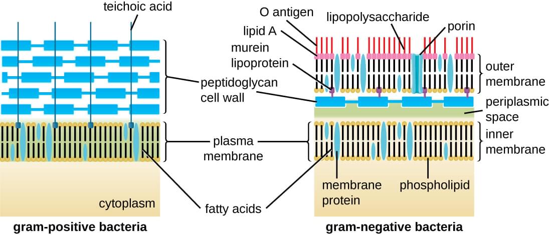
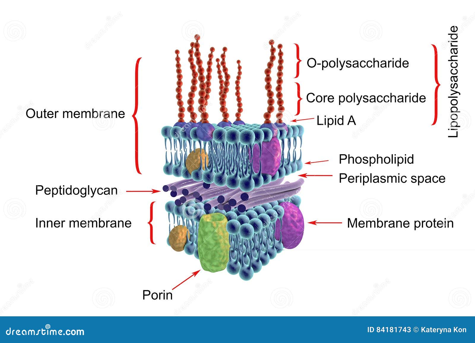
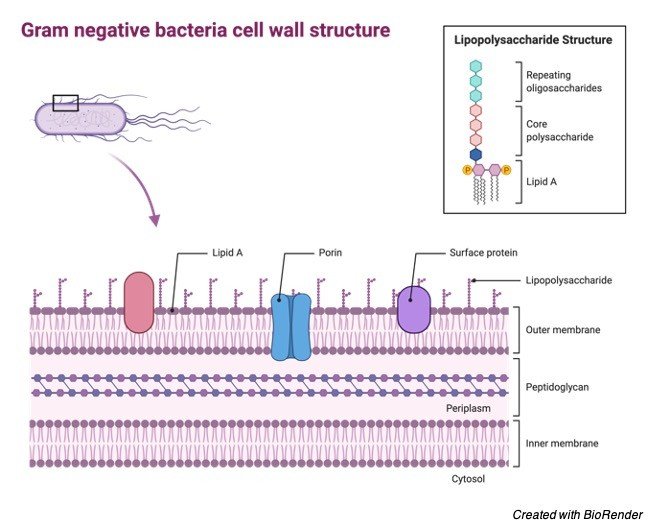
you are gram staining a known sample that contains both gram-positive and gram-negative bacteria after you complete the staining, you realize that you didn't use the alcohol bottle at all which cells would appear purple? gram-positive cells gram-negative cells. 3 MULTIPLE CHOICE OPTIONS. Gram-negative bacteria are bacteria that do not retain the crystal violet stain used in the Gram staining method of bacterial differentiation. They are characterized by their cell envelopes, which are composed of a thin peptidoglycan cell wall sandwiched between an inner cytoplasmic cell membrane and a bacterial outer membrane.. Gram-negative bacteria are found in virtually all environments on ... The cell wall structure of Gram negative bacteria is more complex than that of Gram positive bacteria. Located between the plasma membrane and the thin peptidoglycan layer is a gel-like matrix called periplasmic space. Unlike in Gram positive bacteria, Gram negative bacteria have an outer membrane layer that is external to the peptidoglycan ... The diagram below illustrates the differences in the structure of Gram positive and Gram negative bacteria. The two key features that lead to the differing visualization properties of Gram positive and Gram negative species are the thickness of the peptidoglycan layer and presence or absence of the outer lipid membrane.
Gram Negative Flow Chart. 13a Gram Negative Bacteria Flashcards Quizlet. Flowchart For Identification Of Anaerobic Gram Positive Bacilli 1 Scientific Diagram. Gram Negative Bacteria Positive Microbiology Flowchart Png 1000x702px Gramnegative Anaerobic Anism Area Bacillus. Solved When Scientific Try To Identify And Unknown Bacter Chegg. Differential stains- Gram stain. In contrast to simple stains, differential stains are used to distinguish the difference between bacteria. One of the most well-known differential stains is Gram stain, which differentiates gram-positive and gram-negative bacteria based on the difference in their cell wall structure.. The Gram stain was developed by the Danish bacteriologist Hans Christian Gram ... Flagella occur on both Gram-positive and Gram-negative bacteria, and their presence can be useful in identification. For example, they are found on many species of bacilli but rarely on cocci. In contrast, pili occur almost exclusively on Gram-negative bacteria and are found on only a few Gram-positive organisms (e.g., Corynebacterium renale). Gneg mploc server a schematic flow chart of the experimental design to elish and scientific diagram gram negative bacteria flow chart docx 1 roli dichotomous key oxidase a faecalis denitrificans p aeruginosa fluorescenes course hero dichotomous key flow charts unknown bacteria flow chart gram negative trinity.
Vektor Stok Cell Wall Structure Gramnegative Bacteria Example Tanpa Royalti 1173538834
In segment 13 i.e. Toxin production it is more accurate to write under gram negative bacteria exotoxins and/or endotoxins rather than exotoxins or endotoxins because endotoxins are produced by all gram negative bacteria as it ( the LPS) is an integral part of the outer membrane, so any species produce exotoxins will already produce both.

Gram Negative Bacteria Gram Positive Bacteria Microbiology Flowchart Others Angle Text Infection Png Pngwing
Gram stain and bacterial morphology: Of all the different classification systems, the Gram stain has withstood the test of time. Discovered by H.C. Gram in 1884 it remains an important and useful technique to this day. It allows a large proportion of clinically important bacteria to be classified as either Gram positive or negative based on their
Structure And Function Of Bacterial Cells
The gram-positive bacteria retain the crystal violet colour and stains purple whereas the gram-negative bacteria lose crystal violet and stain red. Thus, the two types of bacteria are distinguished by gram staining. Gram-negative bacteria are more resistant against antibodies because their cell wall is impenetrable.

Structure And Function Of Bacterial Cells
Gram-negative Bacteria. Go to PAGE 2 > Gram-positive Cells. In Gram-positive bacteria, peptidoglycan makes up as much as 90% of the thick cell wall enclosing the plasma membrane. See Page 2 for a diagram of the Gram-negative cell wall and a video on ...

Cell Wall Structures Of Gram Positive And Gram Negative Bacteria And Download Scientific Diagram
The Gram stain is a differential staining technique used to classify & categorize bacteria into two major groups: Gram positive and Gram negative, based on the differences of the chemical and physical properties of the cell wall.
The Outer Membrane Is An Essential Load Bearing Element In Gram Negative Bacteria Nature
Download scientific diagram | Antibiotics used for gram positive and gram negative bacteria with its disc content from publication: Antibiotic sensitivity profile of bacterial pathogens in ...

Bacteria Cell Wall Illustration Gram Positive And Gram Negative Cell Wall Differents Stock Illustration Download Image Now Istock
Gram Negative Bacteria. Like Gram positive bacteria, Gram negative bacteria are also well distributed in different environments across the globe. Whereas E. coli among other Gram negative bacteria can be found in the gastrointestinal tract, a number of species can be found in marine environments.

Gram Positive Vs Gram Negative Biology Dictionary
Mycoplasma are bacteria that have no cell wall and therefore have no definite shape. Outer Membrane: This lipid bilayer is found in Gram negative bacteria and is the location of lipopolysaccharide (LPS) in these bacteria. Gram positive bacteria lack this layer. LPS can be toxic to a host and can stimulate the host's immune system.
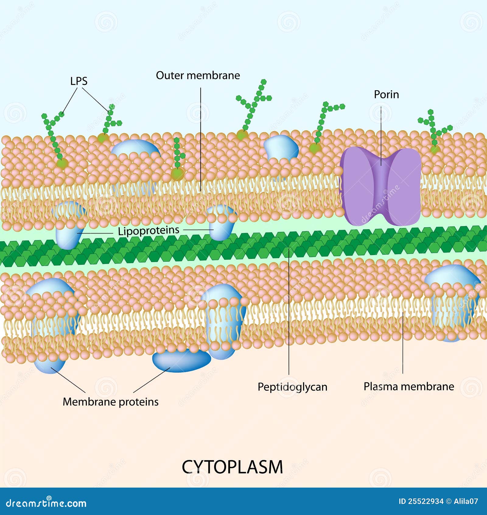
Gram Negative Bacterial Cell Wall Stock Vector Illustration Of Cellular Antibiotics 25522934
4 Bacteria: Cell Walls . It is important to note that not all bacteria have a cell wall.Having said that though, it is also important to note that most bacteria (about 90%) have a cell wall and they typically have one of two types: a gram positive cell wall or a gram negative cell wall.. The two different cell wall types can be identified in the lab by a differential stain known as the Gram stain.

Bacteria Cell Wall Gram Positive And Gram Negative
THE GRAM-NEGATIVE CELL ENVELOPE. After more than a decade of controversy, techniques of electron microscopy were improved to the point in which they finally revealed a clearly layered structure of the Gram-negative cell envelope (Fig. 1) (Glauert and Thornley, 1969).There are three principal layers in the envelope; the outer membrane (OM), the peptidoglycan cell wall, and the cytoplasmic or ...

19 Science And Nature Infographics Ideas Science And Nature Science Science Education
Christian Gram, a Danish Physician in 1884 developed a staining technique to distinguish two types of bacteria. The two categories of bacteria based on gram staining are Gram positive bacteria and Gram negative bacteria. Bacteria are first stained with crystal violet or gentian violet.
Gram Negative Bacteria Microbiology Medbullets Step 1
8. According to Peberdy (1980) the only compound present in the cell walls of both Gram-negative and Gram-positive bacteria is 'peptidoglycan'. The cell walls of Gram-positive bacteria contain up to 95% peptidoglycan and up to 10% teichoic acids. 9. Cytoplasmic membrane is a thin (5-10 nm) layer lining the inner surface of the cell wall.

Graphene Oxide Exhibits Differential Mechanistic Action Towards Gram Positive And Gram Negative Bacteria Sciencedirect
Bacteria can be classified based on shape, mode of nutrition, respiration, the composition of the cell wall, etc. Based on these criteria, bacteria can be classified as bacillus, coccus or vibrio, etc., autotrophic or heterotrophic, aerobic or anaerobic, Gram + or Gram -.
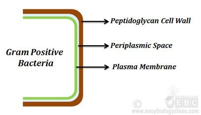
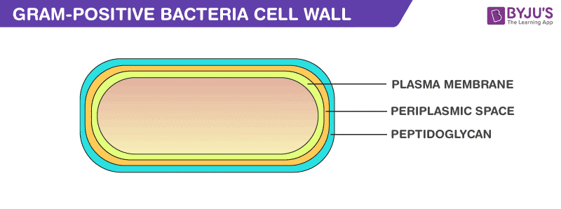
Cell Wall Of Gram Positive Bacteria Easybiologyclass Easy Biology Class
Gram-negative bacteria have a smaller amount of this rigid structure than do gram-positive bacteria. Periplasmic space. an open area between the cell wall and cell membrane in the cell envelopes of bacteria. gram negative bacteria have a more extensive space than do a gram positive bacteria. Cell membrane.
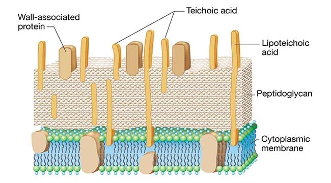
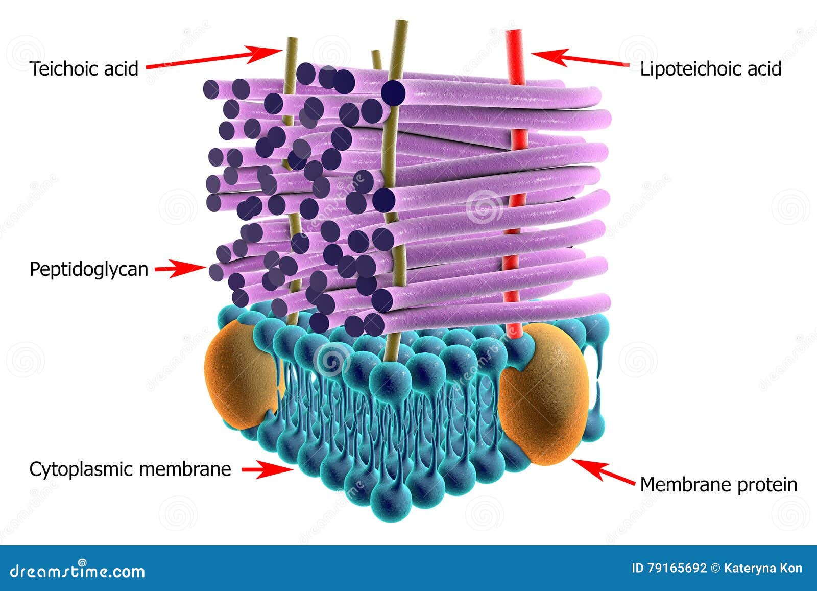
Gram Positive Cell Wall Microbe Notes

Difference Between Gram Positive And Gram Negative Cell Wall Compare The Difference Between Similar Terms

Gram Positive Bacteria Gram Negative Bacteria Gram Stain Coccus Classification Angle Text Png Pngegg

Gram Negative Bacterial Cell Wall Unit 1 Diagram Quizlet

Microbiological Diagram Sample Flagellum Of Gram Negative Bacteria Microbiology Science Education Education

Bacteriophage Derived Endolysins To Target Gram Negative Bacteria Sciencedirect
Gram Positive Bacteria Characteristics And Structure

Schematic Structure Of Gram Positive And Gram Negative Cell Walls Download Scientific Diagram

Hospital Acquired Infections Due To Gram Negative Bacteria Nejm
1
How Can We Change Gram Negative To Gram Positive In Bacteria Quora

Cross Section Of The Envelope Of A Typical Gram Negative Bacterium Lps Download Scientific Diagram
:max_bytes(150000):strip_icc()/gram_negative_bacteria-5b7f308646e0fb002cbcdc5d.jpg)
Gram Positive Vs Gram Negative Bacteria
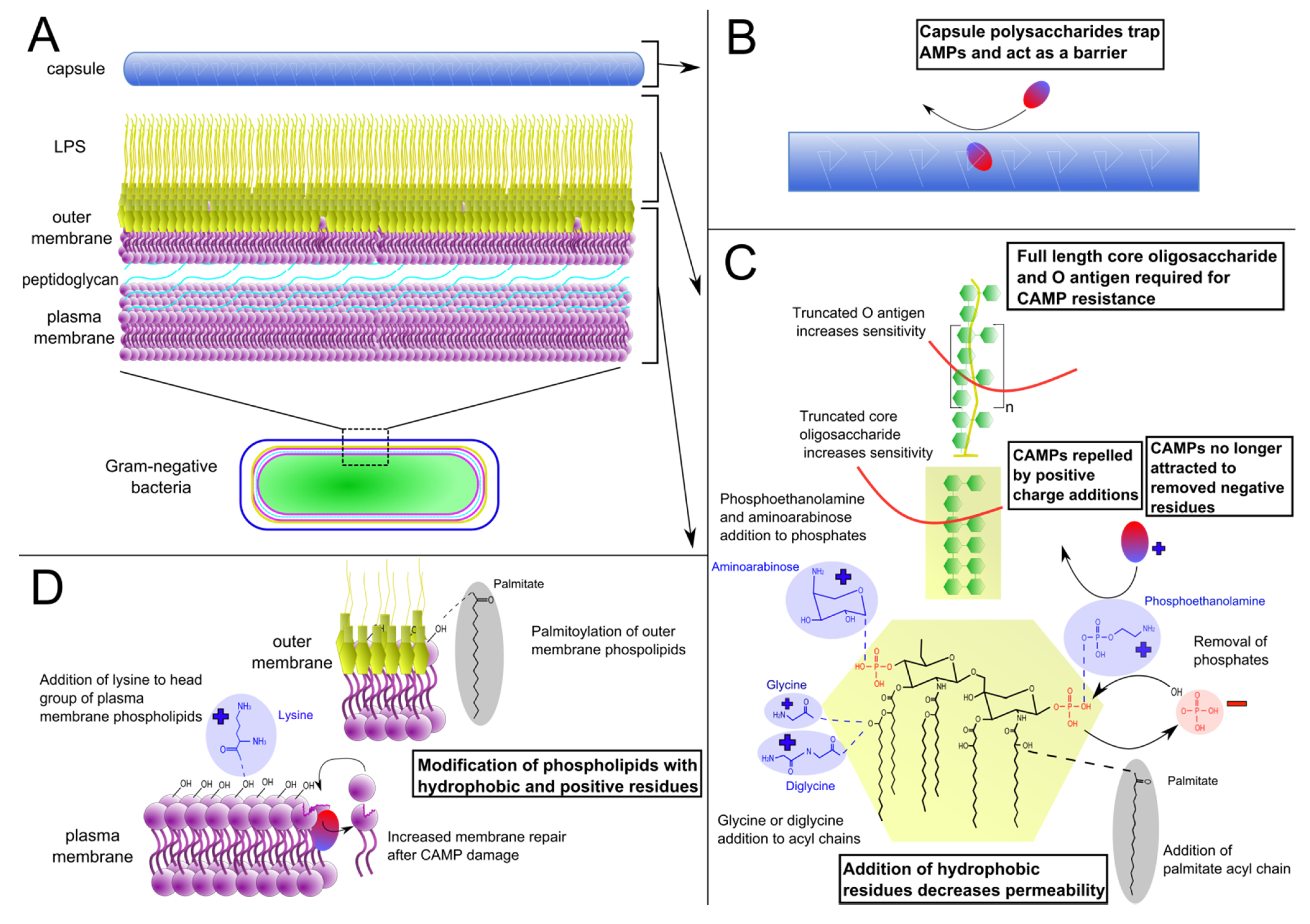
Antibiotics Free Full Text Mechanisms Of Antimicrobial Peptide Resistance In Gram Negative Bacteria
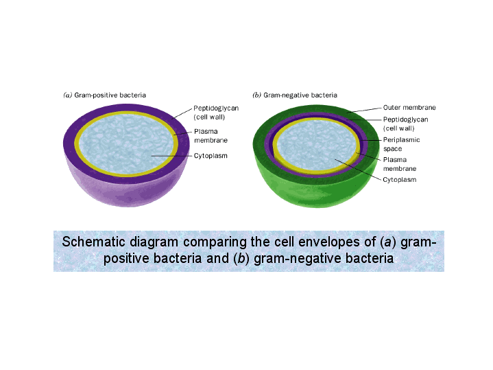
Schematic Diagram Comparing The Cell Envelopes Of A Gram Positive Bacteria And B Gram Negative Bacteria

Structure Of Grampositive Bacteria Cell Wall Stock Photo Download Image Now Istock
Bacterial Cell Wall Targets Identification Creative Biolabs
Structure Of Gram Positive Bacteria Cell Wall Stock Illustration Illustration Of Microbial Muramic 79165692
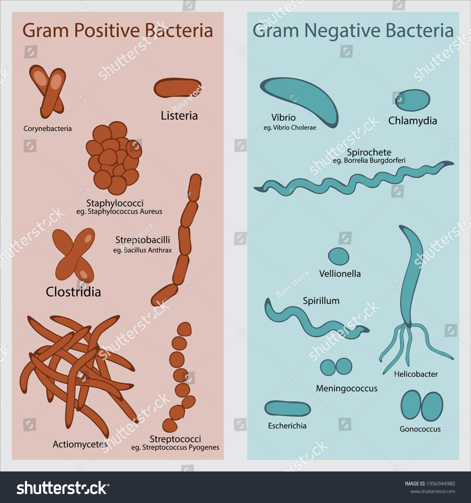
Diagram Showing Example Gram Negative Positive Stock Vector Royalty Free 1956944980
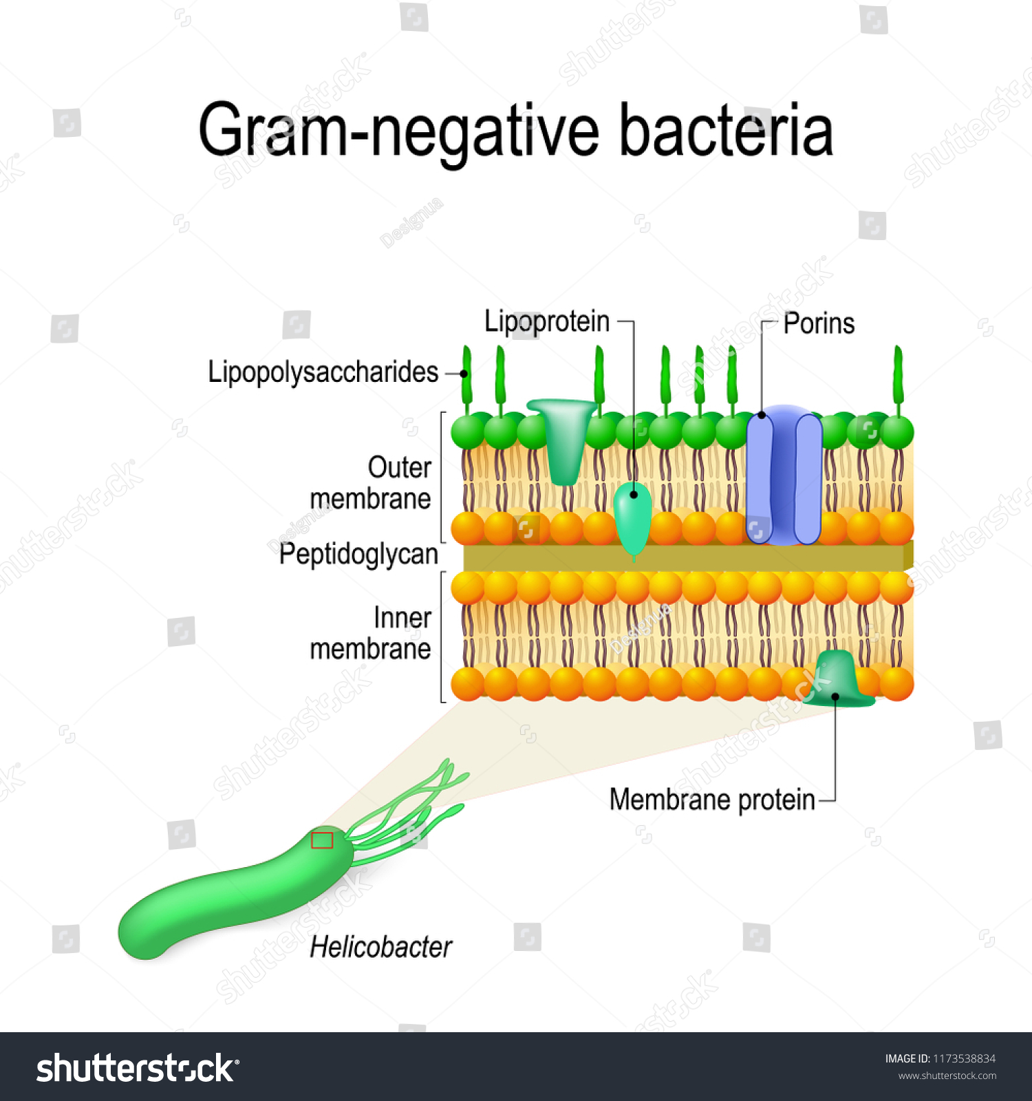
Cell Wall Structure Of Gram Negative Bacteria For Example Helicobacter Vector Diagram For Educational Medical Biological And Science Use Stock Vector Image Art Alamy
Gram Negative Bacteria Characteristics Cell Structure And Diseases
Pathogens Bacteria Viruses Gram Stain Teachmephysiology

Microbiology Notes And Updates Gram Negative Bacteria Cell Wall Structure Follow Us On Instagram For More Pictures Https Www Instagram Com Microbenotes Facebook
Bacterial Cell Structure Cell Wall Gram Positive Bacteria Gram Negative Bacteria Png 696x471px Bacterial Cell Structure
Bacterial Cell Wall Structure Composition And Types Online Biology Notes

Difference Between Gram Positive And Gram Negative Bacteria Youtube

Microbiology Notes And Updates Know More From Https Microbenotes Com Gram Negative Bacteria Facebook
Structure Of Gram Negative Bacteria Cell Wall Stock Illustration Illustration Of Lipopolysaccharide Plasma 84181743

Endotoxin Cell Membrane Bacterial Outer Membrane Gram Negative Bacteria Membrane Transport Protein Angle Text Cell Png Pngwing
Bacterial Cell Wall Structure Gram Positive Vs Gram Negative
0 Response to "44 gram negative bacteria diagram"
Post a Comment