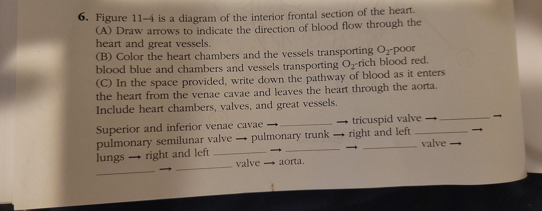
41 figure 11-4 is a diagram of the frontal section of the heart
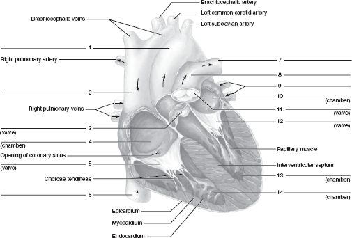
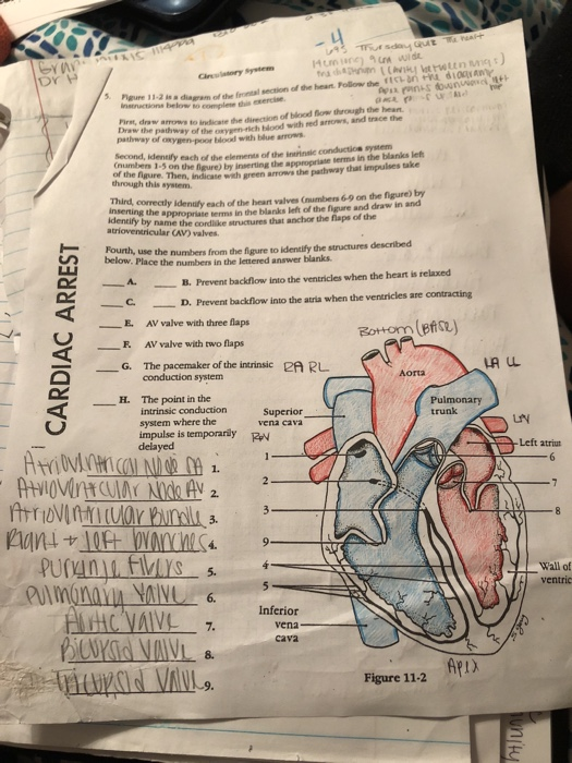
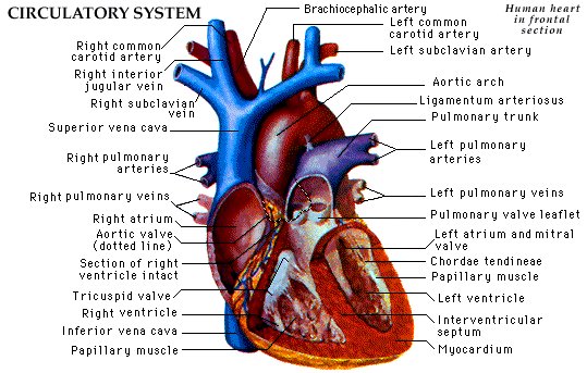
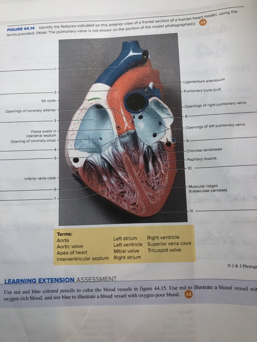
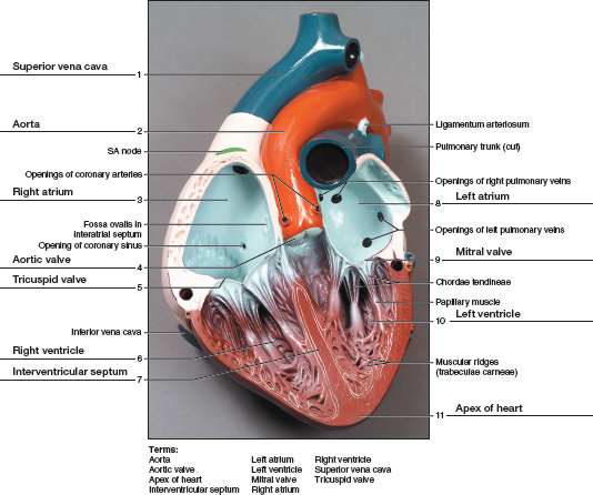
Figure 11-4 is a diagram of the frontal section of the heart. Follow the instructions below to complete this exercise. (a) Correctly identify each of the heart valves (numbers 6-9 on the figure) by inserting the appropriate terms in the blanks left of the figure. (b) Use the numbers from the figure to identify the structures described below. Diagram of Heart. The human heart is the most crucial organ of the human body. It pumps blood from the heart to different parts of the body and back to the heart. The most common heart attack symptoms or warning signs are chest pain, breathlessness, nausea, sweating etc. The diagram of heart is beneficial for Class 10 and 12 and is frequently ...
02.01.2018 · Obesity continues be among the top health concerns across the globe. Despite our failure to contain the high prevalence of obesity, we now have a better understanding of its pathophysiology, and how excess adiposity leads to type 2 diabetes, hypertension, and cardiovascular disease. Lifestyle ...
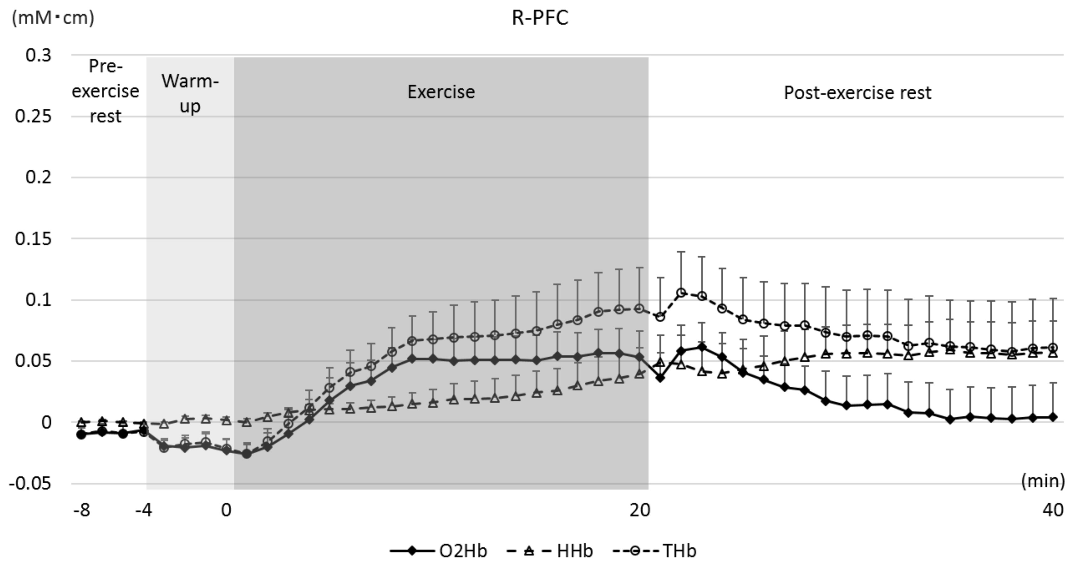
Figure 11-4 is a diagram of the frontal section of the heart
Figure 1.1-2 is a diagram of the frontal section of the heart. Follow the instructions below to complete this exercise. First, draw arrows to indicate the ...1 page Beginning Blender Open Source 3D Modeling, Animation, and Game Design Companion eBook Available Full Color Inside BOOKS FOR PROFESSIONALS BY PROFESSIONALS Beginning Blender: Open Source 3D Modeling, Animation, and Game Design 31 May 2021 — Figure 11-4 is a diagram of the frontal section of the heart instructions ... b the of blood flow through the heart. pathway of o t oxygen-rich.
Figure 11-4 is a diagram of the frontal section of the heart. 6. Figure 11—4 is a diagram of the frontal section of the heart. Follow the instructions below to complete this exercise. First, draw arrows to indicate the direction of blood flow through the heart. Draw the pathway of the oxygen-rich blood with red arrows, and trace the pathway of oxygen-poor blood with blue arrows. 04.05.2020 · Cause of death is predominantly vascular, i.e. heart attacks and strokes as a result of plaques forming within arteries (premature atherosclerosis).|Figure 2: (a) Estimated survival curves for men and women with and without diabetes, and (b) corresponding estimated years of life lost due to diabetes by cause of death. Data from multiple, mainly high-income countries. Source: NEJM The major structures of the cardiovascular system, the heart and blood vessels, ... Figure 11–4 is a diagram of the frontal section of the heart. Follow the.20 pages Figure 11 – 5 Diagram of the frontal section of the heart (pp. 211 – 2112). PowerPoint Presentations: Circulatory System; Cardiovascular System-Marieb 7thEd ...
Figure 1 is a simple diagram of normal skin structure. It also indicates the major cell types that are normally found in each compartment. Broadly speaking, there are two large compartments—the avascular epidermis and the vascular dermis—with many cell types distributed in a connective tissue matrix, largely created by fibroblasts. Enlarge Figure 1. Schematic representation of normal skin ... The Alhambra (Figure 6-33) ... The question of how we decide on such a controversial issue is at the heart of the humanities, and some observers have pointed to Mary Wollstonecraft Shelley’s famous novel Fran-kenstein, Or the Modern Prometheus, which in some ways enacts the conflict among these values. These examples demonstrate that the discoveries of scientists often have tre-mendous ... Figure 11-4 is a diagram of the frontal section of the heart. Follow the instructions below to complete this exercise. (a) Correctly identify each of the heart valves (numbers 6-9 on the figure) by inserting the appropriate terms in the blanks left of the figure. (b) Use the numbers from the figure to identify the structures described below. Um. Page 10. Chapter 11 The Cardiovascular System. 181. 6. Figure 11–4 is a diagram of the frontal section of the heart. Follow the instructions below to ...18 pages
The heart is upside down at this point, with the ventri-cle above the two incoming vessels. Around week 5, the heart curves back on itself in an “S” turn, creating the familiar anatomy, with the atria on top (Fig-ure 11.3). The adult heart is shown in . Figure 11.4. Note the thick ventricular walls, especially in the left ventricle. Chapter 11 The Cardiovascular System 181 6. Figure 11-4 is a diagram of the frontal section of the heart. Follow the instructions below to complete this exercise. First, draw arrows to indicate the direction of blood flow through the heart. Draw the pathway of the oxygen-rich blood with red arrows, and trace the pathway of oxygen-poor blood ... 26.03.2018 · Following this procedure, we found 1824 publications and, after applying the selection criteria, the total number of relevant publications was reduced to 12 (see Results section). To document the literature search process, we used the Preferred Reporting Items for Systematic Reviews and Meta-Analyses (PRISMA): Figure 1 shows the process diagram followed to select the included studies. Figure 11-4 is a diagram of the frontal section of the heart. Follow the instructions below to complete this exercise. First, draw arrows to indicate the ...
Applied Sciences Free Full Text Relationship Between The Difference In Oxygenated Hemoglobin Concentration Changes In The Left And Right Prefrontal Cortex And Cognitive Function During Moderate Intensity Aerobic Exercise Html
Academia.edu is a platform for academics to share research papers.

The Pathophysiology Of Cigarette Smoking And Cardiovascular Disease An Update Journal Of The American College Of Cardiology
6. Figure 11—4 is a diagram of the frontal section of the heart. Follow the instructions below to complete this exercise. The colored arrows indicate the flow of oxygenated (red) and deoxygenated (blue) blood through the heart. First, identify each of the elements of the intrinsic conduction system (red text boxes) by inserting the appropriate terms in the blanks left of the figure.
Solved 6 Figure 11 4 Is A Diagram Of The Interior Frontal Chegg Com
Figure 14.5.3 – Phineas Gage: The victim of an accident while working on a railroad in 1848, Phineas Gage had a large iron rod impaled through the prefrontal cortex of his frontal lobe. After the accident, his personality appeared to change, but he eventually learned to cope with the trauma and lived as a coach driver even after such a traumatic event. (credit b: John M. Harlow, MD)

Chapter 20 The Cardiovascular System The Heart Ppt Download
The first major branch off of the aorta and the major artery to the forelimbs and head. Superior Vena Cava. receives blood from the head and arms and chest and empties into the right atrium of the heart. Ascending Aorta. the ascending part of the aorta as it emerges from the left ventricle. Pulmonary Semilunar Valve.
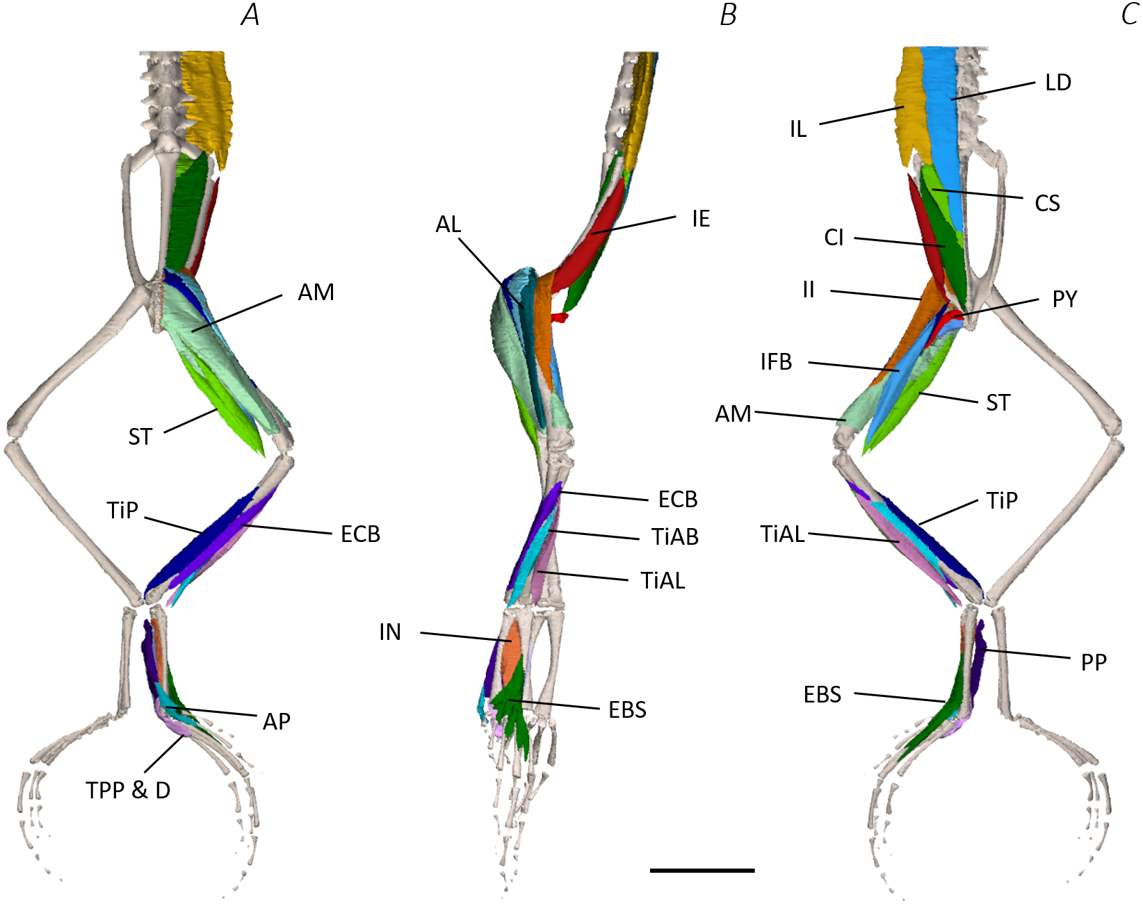
Digital Dissection Of The Pelvis And Hindlimb Of The Red Legged Running Frog Phlyctimantis Maculatus Using Diffusible Iodine Contrast Enhanced Computed Microtomography Dice Mct Peerj
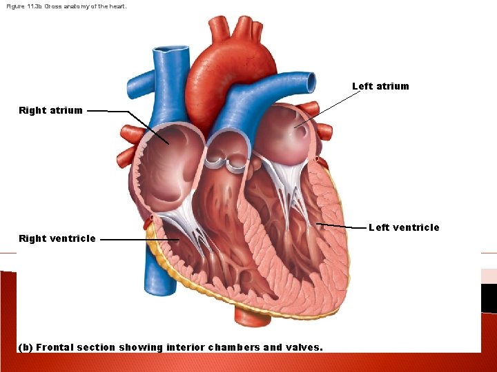
of the blood vessels leaving and entering the heart. (Figure 11.3 shows two views of the heart—an exter-nal anterior view and a frontal section. As the ana-tomical areas of the heart are described in the next section, keep referring to Figure 11.3 to locate each of the heart structures or regions.) Chambers and Associated Great Vessels
Chapter 44 Solutions Laboratory Manual For Human Anatomy Physiology Fetal Pig Version 2nd Edition Chegg Com
Anatomy and Physiology questions and answers. 6. Figure 11-4 is a diagram of the interior frontal section of the heart. (A) Draw arrows to indicate the direction of blood flow through the heart and great vessels. (B) Color the heart chambers and the vessels transporting O2-poor blood blue and chambers and vessels transporting O2-rich blood red.
Applied Sciences Free Full Text Flavor Chemistry Of Virgin Olive Oil An Overview Html
07.11.2021 · The heart is (7) to the vertebral column (spine) and (8) to the lungs. 9/5/2017 Laboratory Manual for Anatomy and Physiology PRINTED BY: After completing this exercise, you should be able to: 1 Describe the anatomical position 2 Use anatomical and directional terms correctly 3 Identify body planes Human Anatomy and Physiology The #1 best-selling Human Anatomy & Physiology …
1
Figure 7.3 The cellular structure of a leaf as seen in a transverse section: Z in Figure 7.2 • Waxy cuticles on the outside of both the upper and lower epidermis are waterproof so they can prevent leaves losing water that is needed for photosynthesis. • Stomatal pores, which are present throughout the lower epidermis, allow carbon dioxide to diffuse into the leaf and oxygen to diffuse out ...
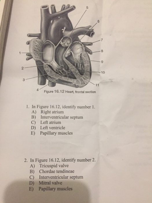
Solved 8 9 10 3 Figure 16 12 Heart Frontal Section 1 In Chegg Com
31 May 2021 — Figure 11-4 is a diagram of the frontal section of the heart instructions ... b the of blood flow through the heart. pathway of o t oxygen-rich.

Frontal Section Anatomy Of Heart Diagram Quizlet
Beginning Blender Open Source 3D Modeling, Animation, and Game Design Companion eBook Available Full Color Inside BOOKS FOR PROFESSIONALS BY PROFESSIONALS Beginning Blender: Open Source 3D Modeling, Animation, and Game Design

Chapter 20 The Cardiovascular System The Heart Ppt Download
Figure 1.1-2 is a diagram of the frontal section of the heart. Follow the instructions below to complete this exercise. First, draw arrows to indicate the ...1 page
2
1

Anatomy Of Heart Interior Frontal Section High Res Vector Graphic Getty Images
The Cardiovascular System The Heart And Circulation Pages
2

Focal Venous Hypertension As A Pathophysiologic Mechanism For Tissue Hypertrophy Port Wine Stains The Sturge Weber Syndrome And Related Disorders Proof Of Concept With Novel Hypothesis For Underlying Etiological Cause An American Ophthalmological
Solved Ani 10 Dr Nnana Tdiagram Of The Frontal Section Of Chegg Com
Ampullary High Resolution Stock Photography And Images Alamy

Anterior Heart Frontal Section Part Two Diagram Quizlet
Digital Dissection Of The Pelvis And Hindlimb Of The Red Legged Running Frog Phlyctimantis Maculatus Using Diffusible Iodine Contrast Enhanced Computed Microtomography Dice Mct Peerj

Frontal Section Through The Heart Diagram Quizlet
2
Frontal Section View Of Heart

Frontal Section Of Heart Diagram Quizlet
Solved Figure 44 14 Identify The Features Indicated On This Chegg Com

3 Sketch Of Superior Frontal Section Showing Scalp Skull And Meninges Download Scientific Diagram

Internal Features Of The Heart Frontal Section Diagram Quizlet

Chronological And Morphological Study Of Heart Development In The Rat Marcela 2012 The Anatomical Record Wiley Online Library

Short Communication Effect Of Age At Group Housing On Behavior Cortisol Health And Leukocyte Differential Counts Of Neonatal Bull Dairy Calves Journal Of Dairy Science
Chapter 44 Solutions Laboratory Manual For Human Anatomy Physiology Cat Version 3rd Edition Chegg Com

Science Poem By Andrew Nealy

Ijerph Free Full Text Education Technology In Orthodontics And Paediatric Dentistry During The Covid 19 Pandemic A Systematic Review Html

Figure 35 3 Frontal Section Of Human Heart Diagram Quizlet

The Heart Ppt Video Online Download

Introduction To Cardiovascular System Ppt Video Online Download
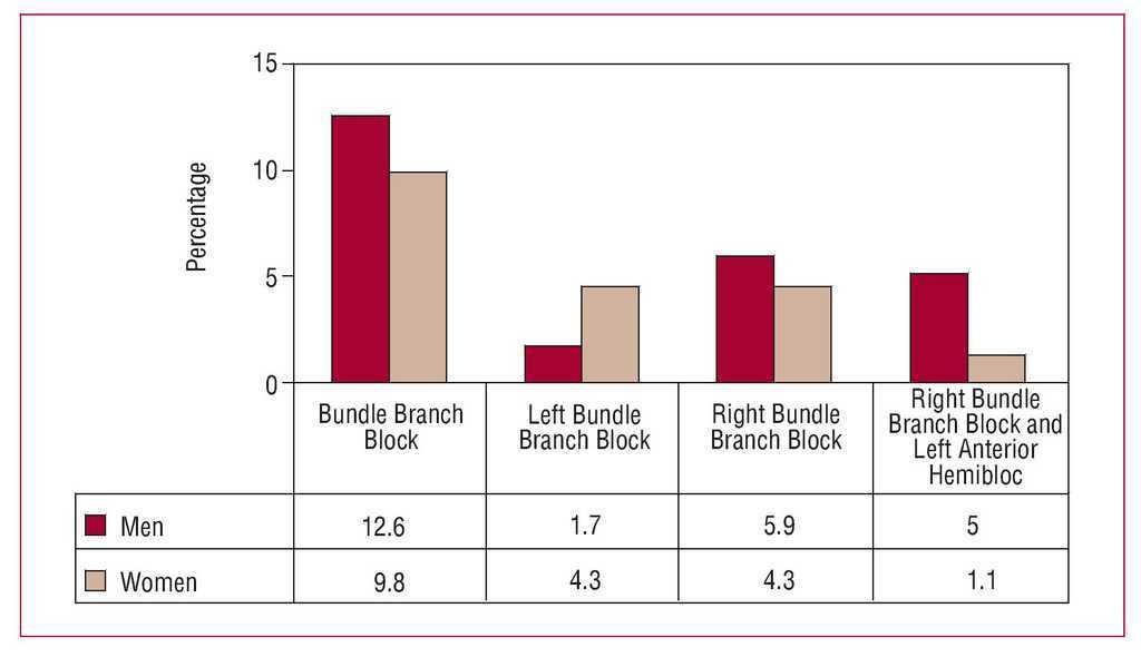
Prevalence Of And Predisposing Factors For Bundle Branch Block In Patients Starting Dialysis Revista Espanola De Cardiologia
2
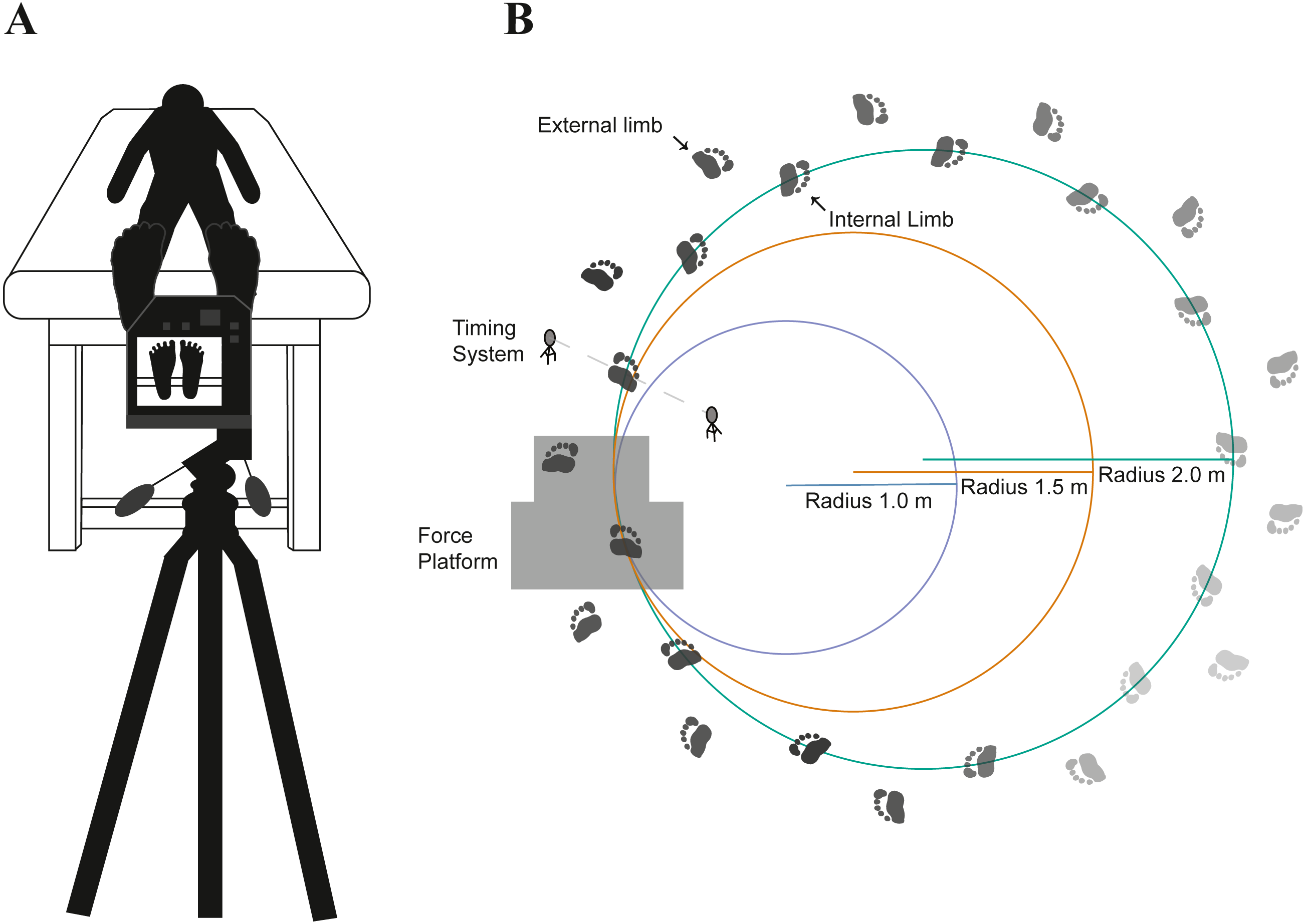
Association Between Foot Thermal Responses And Shear Forces During Turning Gait In Young Adults Peerj

Third Universal Definition Of Myocardial Infarction Circulation

Frontal Section Of The Heart A Diagram Quizlet
0 Response to "41 figure 11-4 is a diagram of the frontal section of the heart"
Post a Comment