45 nerves in the head diagram
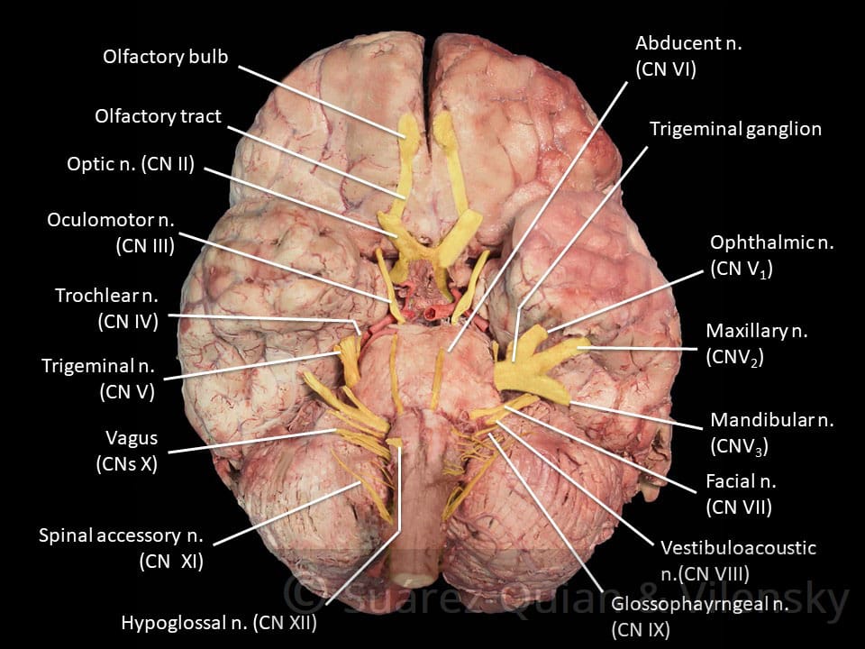
12 Cranial Nerves: Functions & Diagram of Locations ... The trigeminal nerve is the largest of the cranial nerves and can be further divided into three divisions: ophthalmic, maxillary, and mandibular. The ophthalmic division sends information from the scalp, forehead, and upper eye lids (the upper parts of the head) and is sensory in modality, of the general somatic sensory variety. Nerves of Head and Neck - Earth's Lab Major nerves supplying the Head are: Cranial Nerve Nerves of Submandibular Region Mandibular Nerve Chorda Tympani Nerve Otic Ganglion Cranial Nerves Cranial nerves are the 12 nerves that emerge directly from the brain.
Nerves of the Head and Neck | Interactive Anatomy Guide The nerves of the head and neck include the most vital and important organs of the nervous system — the brain and spinal cord — as well as the organs of the special senses. In addition, in this region we also find the major cranial and spinal nerves that connect the central nervous system to the organs, skin, and muscles of the head and neck.
:max_bytes(150000):strip_icc()/GettyImages-141483691-4cc225237a5945f8ab949d936f52c48e.jpg)
Nerves in the head diagram
Head anatomy: Muscles, glands, arteries and nerves | Kenhub The nerves in the head originate from three main sources: Trigeminal nerve (CN V) Facial nerve (CN VII) Cervical plexus The trigeminal nerve supplies sensory innervation to the forehead and cheek regions of the face via its ophthalmic and maxillary branches. What are the Nerves in the Neck? (with pictures) A diagram showing nerves in the head and neck. Nerves in the neck, medically referred to as the cervical spine, help transmit information along the pathways of the central and peripheral nervous system, including sensory and motor skills processes. The cervical spine consists of eight different sets of nerves. Body Anatomy: Upper Extremity Nerves | The Hand Society Peripheral (outside of the central nervous system) nerves are tubes that are special in their ability to transmit electric impulses along their length and into or away from the central nervous system. Nerves have specialized receptors for different inputs like hot, cold, sharp and vibration. Smaller nerves are grouped into larger rope-like groups that travel up and down the body.
Nerves in the head diagram. Nerves of Human Heart and their Action (With Diagram ... ADVERTISEMENTS: The regulation of the heart is effected through the afferent (centripetal) and efferent (centrifugal) nerves of the heart (Fig. 7.79). The afferent nerves are: ADVERTISEMENTS: i. From the heart through the vagus nerve and from the aortic arch, the aortic nerve. ii. From the heart through the inferior cervical and first four thoracic ganglia […] Nerves of the Face, Head, Neck, and Chest Diagram | Quizlet Start studying Nerves of the Face, Head, Neck, and Chest. Learn vocabulary, terms, and more with flashcards, games, and other study tools. Neck Anatomy Pictures Bones, Muscles, Nerves Nerves within the Cervical Spine: Neck Anatomy Nerves Picture There are 8 spinal nerves that originate from the cervical spine. The majority of these nerves control the functions of the upper extremities and allow you to feel your arms, shoulder, and back of your head. Each nerve provides sensation to a specific area of the body called a dermatome. Cervical Spinal Nerves - Spine-health C1, C2, and C3 (the first three cervical nerves) help control the head and neck, including movements forward, backward, and to the sides. 1 The C2 dermatome handles sensation for the upper part of the head, and the C3 dermatome covers the side of the face and back of the head. 2 (C1 does not have a dermatome.)
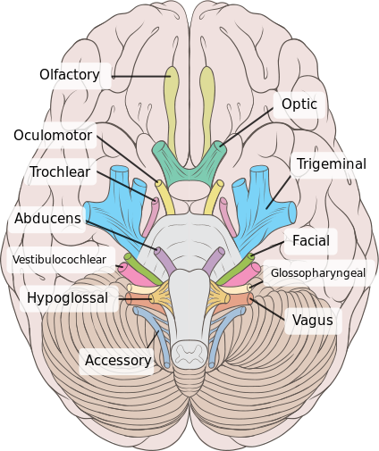
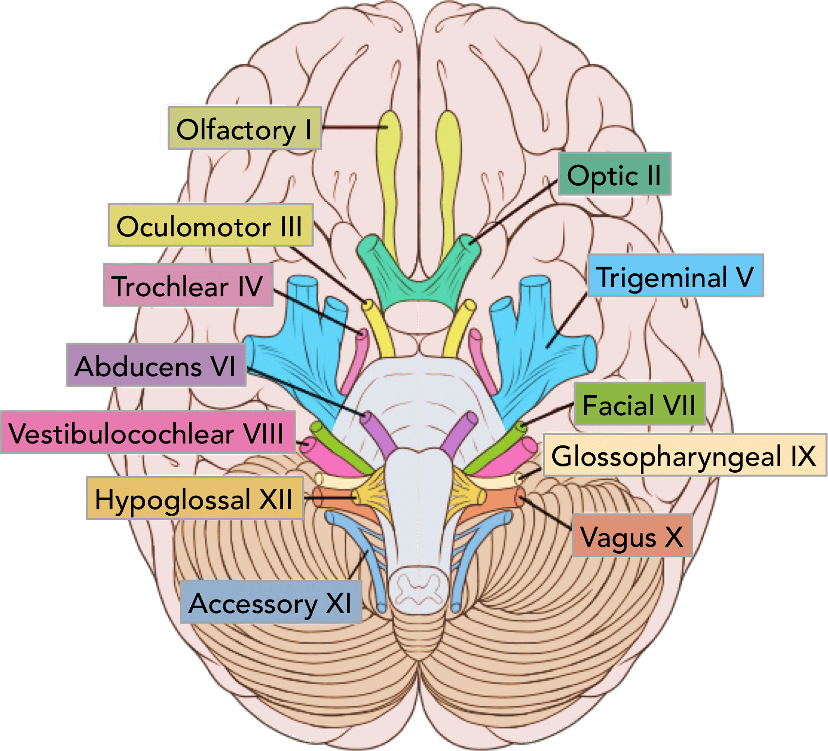
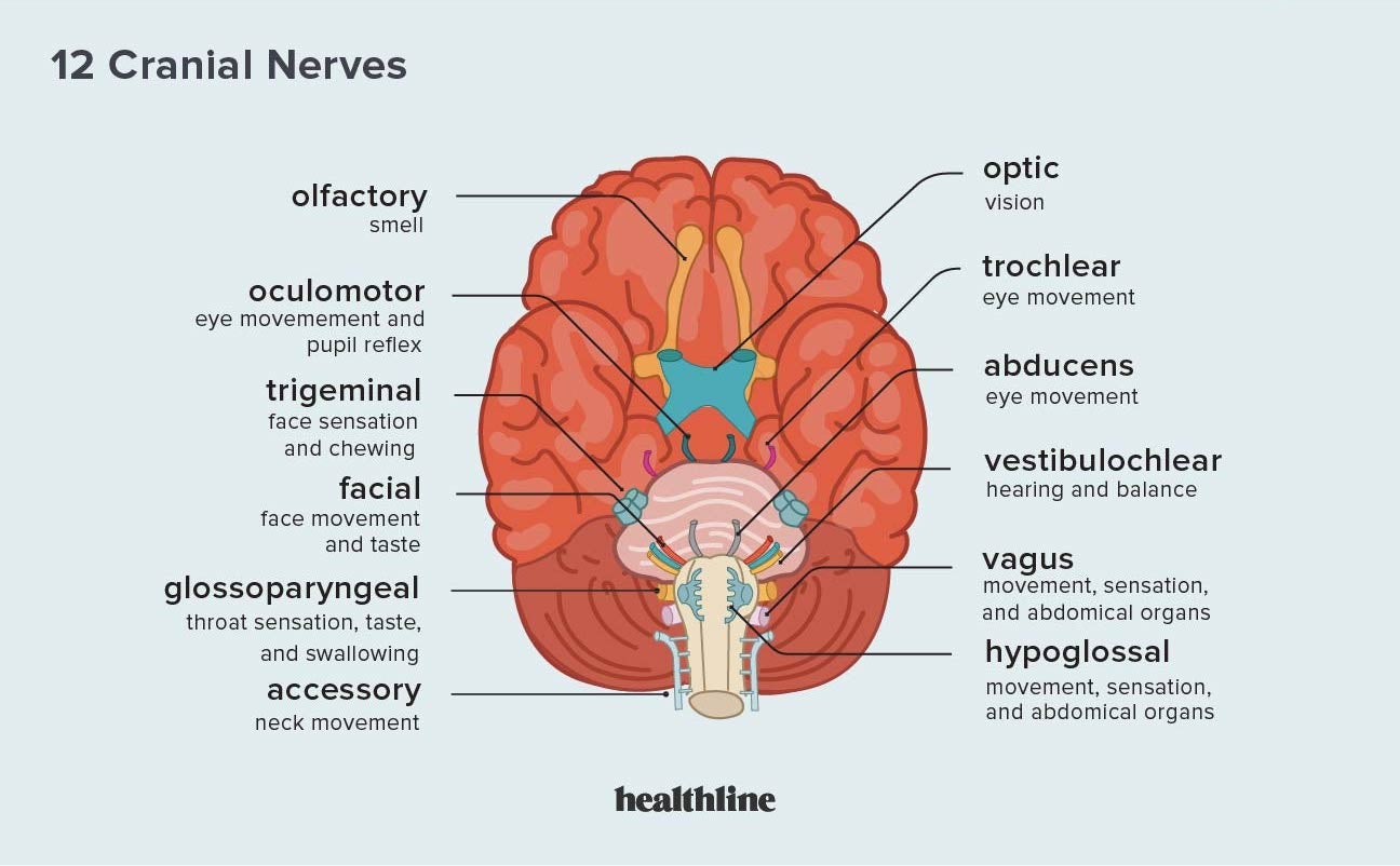
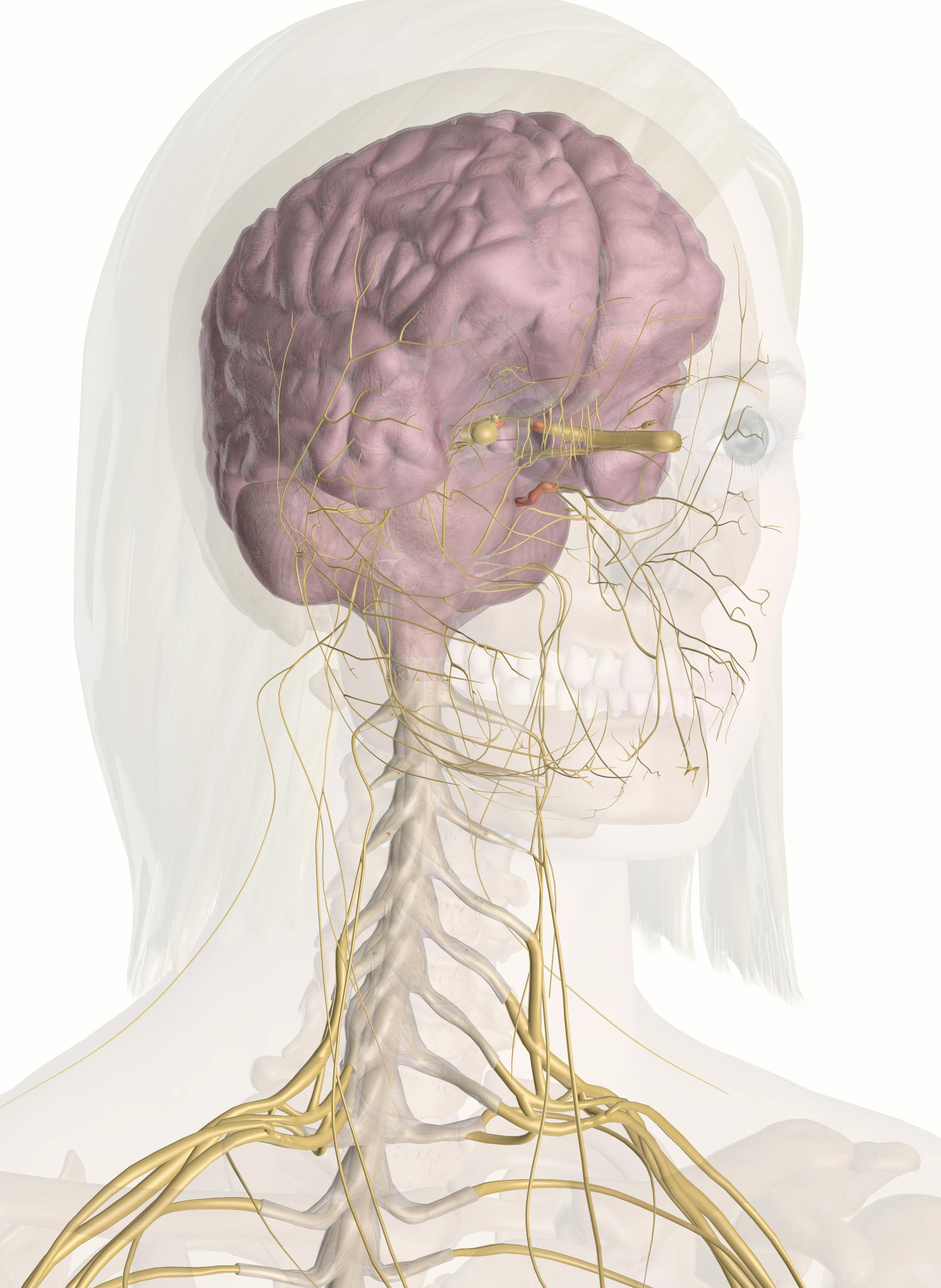
Cranial Nerve Anatomy / Cranial nerves | Iowa Head and ... A thorough understanding of the trigeminal nerve anatomy may be utilized for very effective local anesthesic blocks used in many procedures of the head and neck including nasal fracture reduction, laceration repair, excision of facial lesions, local flap repair, septal hematoma drainage, dental procedures, and intraoral procedures. Major arteries, veins and nerves of the body: Anatomy | Kenhub The 12 pairs of the cranial nerves originate from the brain. They are the: olfactory (CN I), optic (CN II), oculomotor (CN III), trochlear (CN IV), trigeminal (CN V), abducens (CN VI), facial (CN VII), vestibulocochlear (CN VIII), glossopharyngeal (CN IX), vagus (CN X), accessory (CN XI), and hypoglossal nerves (CN XII). Instant Anatomy - Head and Neck - Nerves - Autonomic Instant anatomy is a specialised web site for you to learn all about human anatomy of the body with diagrams, podcasts and revision questions The Nerves at the Back of Your Head and Headaches - Regenexx In red are the deep muscles that control your head on your neck. In yellow, you see the following nerves: GON - Greater occipital nerve - This along with the TON originate from the C2 spinal nerve. The GON comes exits alongside the upper trapezius (not shown as that muscle is more superficial than the deep ones illustrated above).
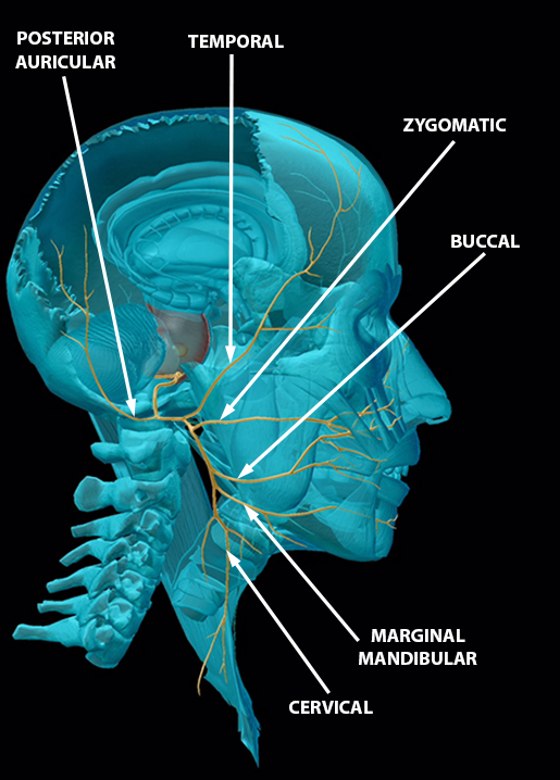
Facial Nerve: Anatomy, Function, and Treatment Six of the facial nerve branches control facial movement. The temporal nerve controls the frontalis muscle. The zygomatic nerve controls the orbicularis oculi. The buccal nerve controls the buccinator and orbucularis oris muscles. The mandibular nerve controls the mentalis muscle. Anatomy, Head and Neck, Occipital Nerves - StatPearls ... The occipital nerves are a group of nerves that arise from the C2 and C3 spinal nerves.[1][2] They innervate the posterior scalp up as far as the vertex and other structures as well, such as the ear.[2] There are three major occipital nerves in the human body: the greater occipital nerve (GON), the lesser (or small) occipital nerve (LON), and the third (or least) occipital nerve (TON). Anatomy, Head and Neck, Cervical Nerves - StatPearls ... Cervical nerves are spinal nerves that arise from the cervical region of the spinal cord. These nerves conduct motor and sensory information via efferent and afferent fibers, respectively, to and from the central nervous system. While classified as peripheral nerves, the motor cell body resides in the anterior horn of the spinal cord. There are eight pairs of cervical nerves, denoted C1 to C8 ... Head Arteries & Nerves Anatomy, Function & Diagram | Body Maps There are 12 pairs of major nerves called cranial nerves that serve both sides of the body.All but two pairs—olfactory and optic—emerge from the brain stem. These two pairs arise from the ...
Cranial Nerves: Anatomy, Function, and Treatment
Nervous System: Explore the Nerves with Interactive ... Thus, there are 8 pairs of cervical nerves, 12 pairs of thoracic nerves, 5 pairs of lumbar nerves, 5 pairs of sacral nerves, and 1 pair of coccygeal nerves. Each spinal nerve exits from the spinal cord through the intervertebral foramen between a pair of vertebrae or between the C1 vertebra and the occipital bone of the skull. Meninges
Cranial Nerve Anatomy / Cranial nerves | Iowa Head and Neck ...
What are the 12 cranial nerves? Functions and diagram The functions of the cranial nerves are sensory, motor, or both: Sensory cranial nerves help a person to see, smell, and hear. Motor cranial nerves help control muscle movements in the head and neck.

Brain regions and nerve Info Graphic #MSEducation ...
Illustrations and diagrams of the 12 pairs of cranial ... Oculomotor nerve [III] , Trochlear nerve [IV] , Abducent nerve; Abducens nerve [VI] : Anatomy atlas. The dermatomes of the trigeminal nerve (V) are represented on a diagram of the face. The branches of the trigeminal nerve (V) are represented in three different diagrams. The ophthalmic nerve (V1) in the orbital cavity with its main branches ...

12 Cranial Nerves: Functions & Diagram of Locations | Simply ...
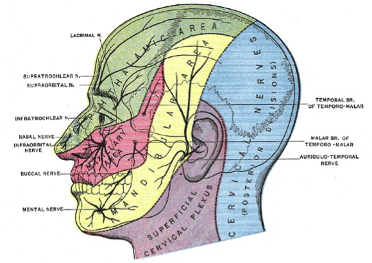
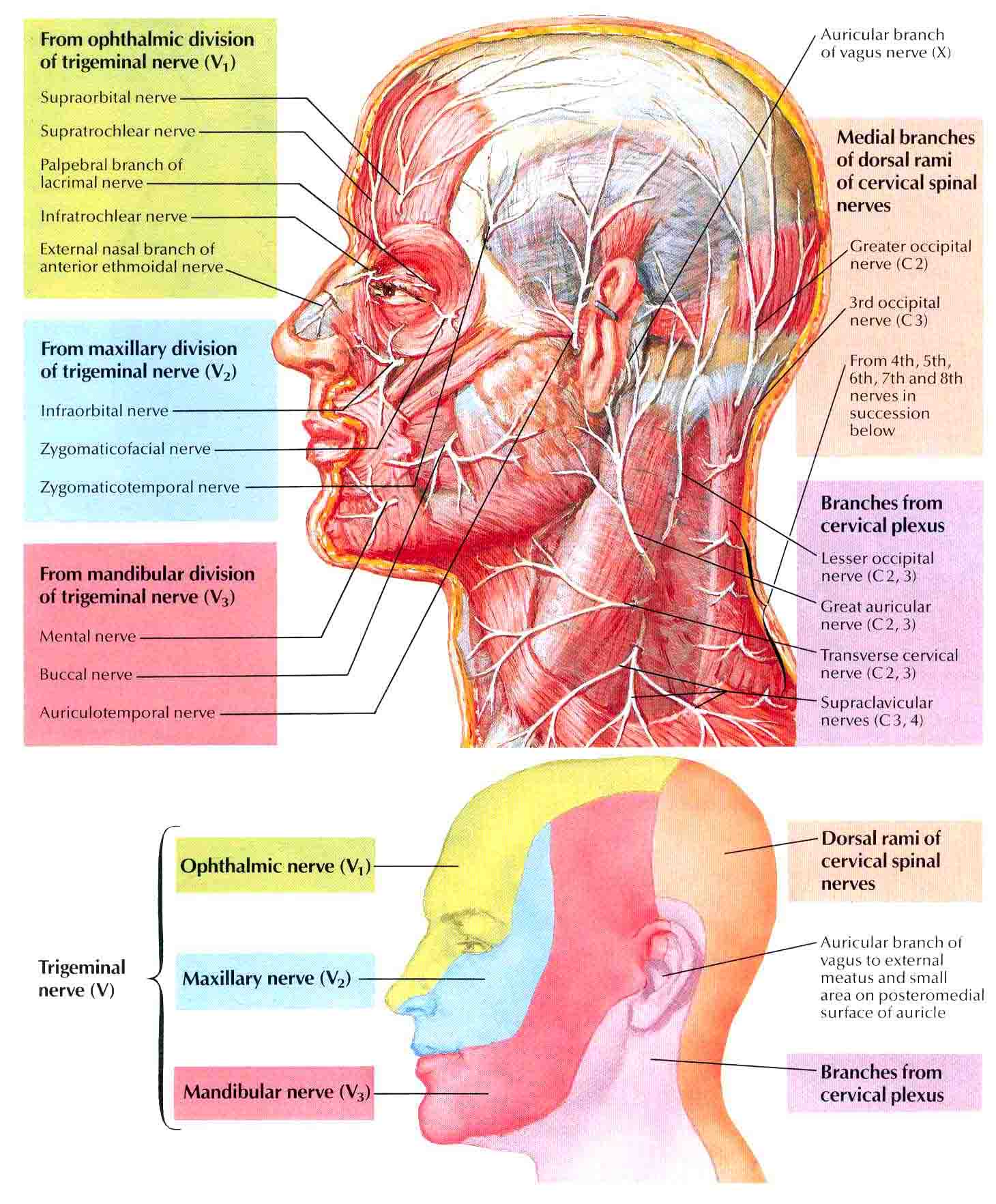
Nerves of the Head - TeachMeAnatomy The nerves of the head include the sympathetic and parasympathetic innervation to the head and neck, as well as the three branches of the trigeminal nerve: ophthalmic, maxillary and mandibular.. The sympathetic innervation begins in the spinal cord.Nerve fibres exit the spinal cord and enter the sympathetic chain, which is composed of superior, middle and inferior cervical ganglion.

Cranial nerves - Wikipedia
Diagram Of The Cutaneous Nerves Of The Head And Neck. News ... Diagram Of The Cutaneous Nerves Of The Head And Neck. (Photo By Encyclopaedia Britannica/UIG Via Getty Images) Diagram Of The Cutaneous Nerves Of The Head And Neck. : News Photo. You have view only access under this Premium Access agreement. Contact your company to license this image.
Summary of the Cranial Nerves - TeachMeAnatomy
Human Nervous System - Diagram - How It Works | Live Science The Cranial Nervous System nerves connect the brain to the eyes, mouth, ears and other parts of the head. The Autonomic Nervous System nerves connect the central nervous system to the lungs, heart ...

Inferior View of Brain: & cranial nerves Diagram | Quizlet
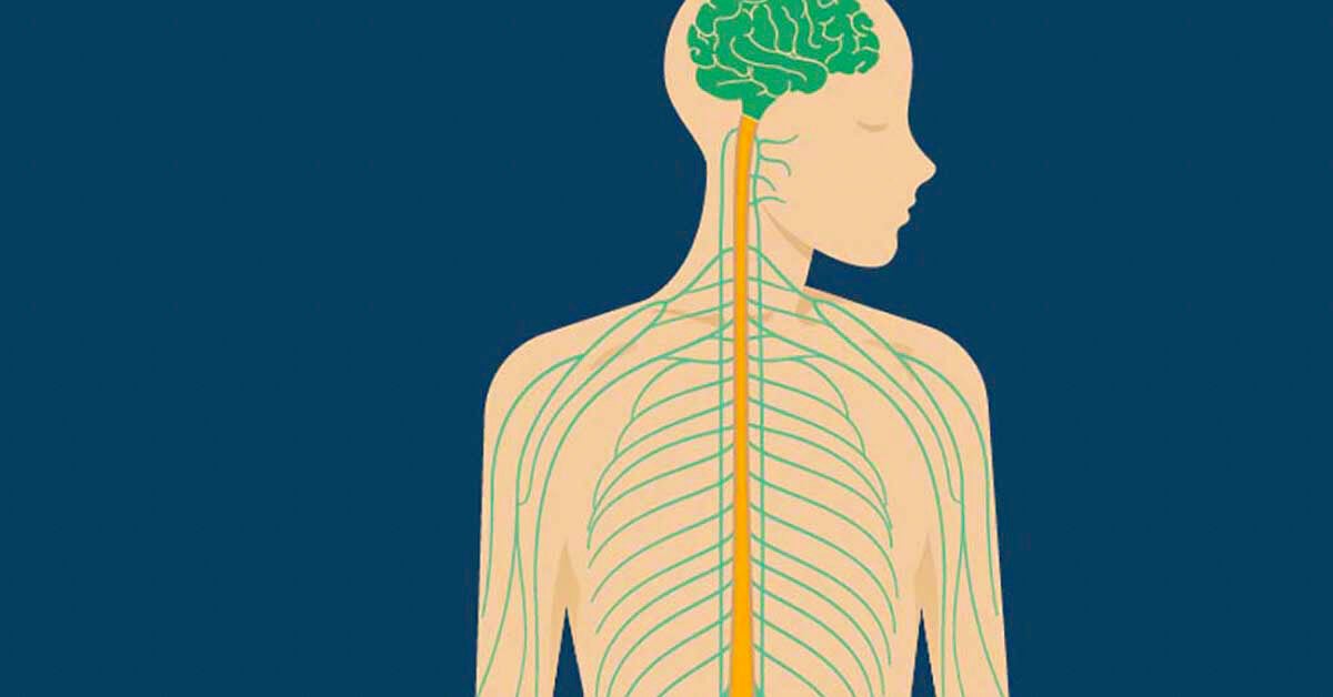
Head and the Nervous System Diagram | Body Maps The nervous system consists of the brain, spinal cord, and nerves. This is the way the body communicates with the brain and vice versa. The nervous system is divided into two key parts: C entral ...
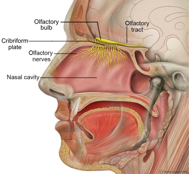
File:Head Olfactory Nerve Labeled.png - Wikimedia Commons
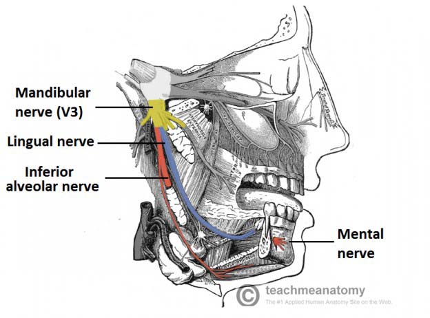
Mandibular Nerve: Anatomy, Function, and Treatment The mandibular nerve, which plays an important role in moving your mouth, splits off from the trigeminal nerve to connect with the lower jaw. It plays both a motor and sensory role in your head as well as interacting with fibers of other cranial nerves.It's the largest of the three branches of the trigeminal nerve, which is the fifth cranial nerve.
Human Nervous System Diagram Stock Illustrations – 2,471 ...
Nerve Supply Of The Jaws And Teeth - Dental Anatomy Twelve pairs of cranial nerves arise in the brain and give off branches to the structures of the head and face. These nerves leave the cranial cavity through foramina in the base of the cranium. The fifth cranial nerve (the trigeminal nerve) is the largest of the twelve pairs. See figure 2-13.

What Is Central Nervous System? Definition, Function & Parts
Occipital Neuralgia - Causes, Symptoms, Diagnosis and ... Occipital Neuralgia is a condition in which the occipital nerves, the nerves that run through the scalp, are injured or inflamed.This causes headaches that feel like severe piercing, throbbing or shock-like pain in the upper neck, back of the head or behind the ears.
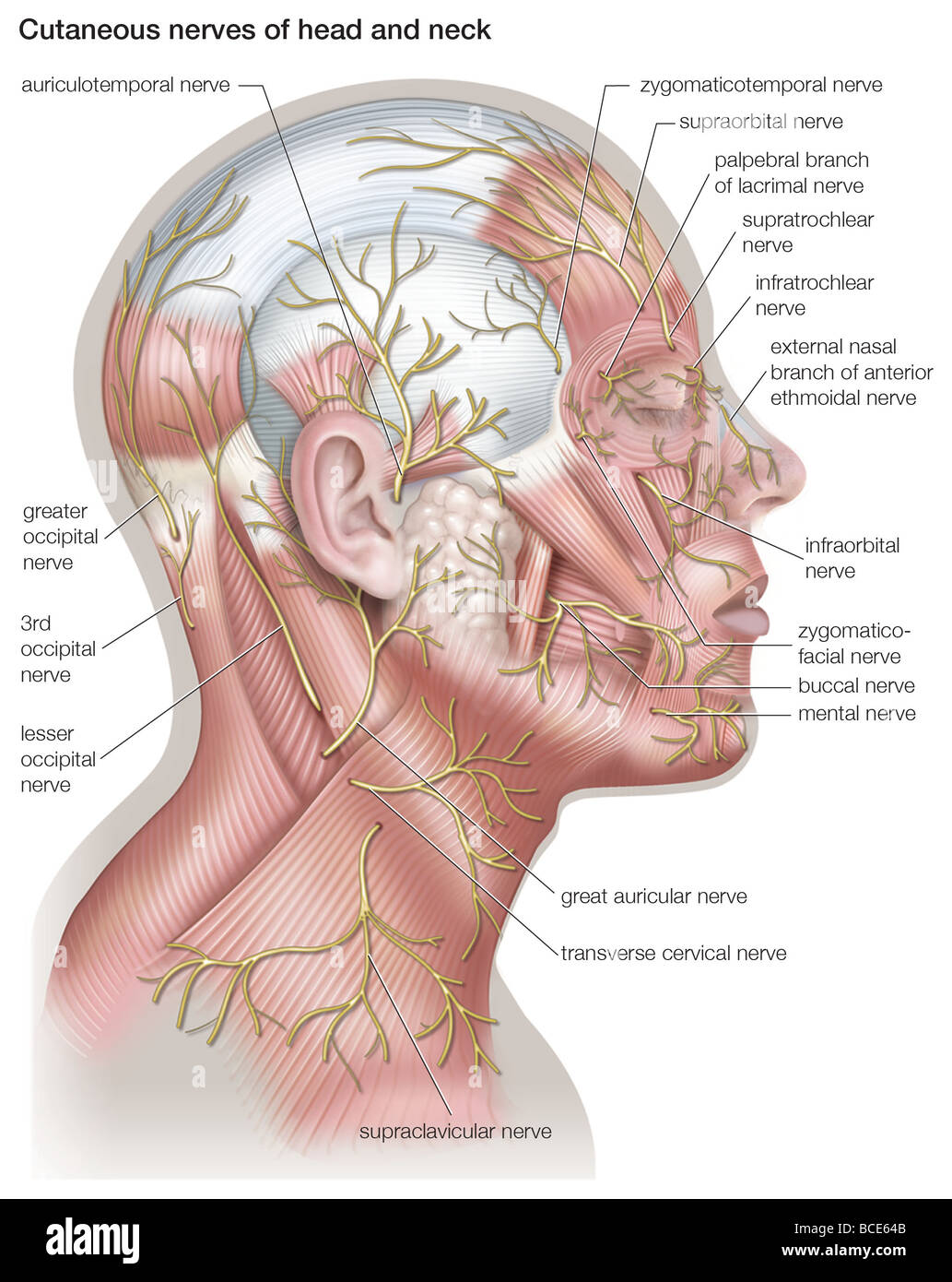
Diagram of the cutaneous nerves of the head and neck Stock ...
Body Anatomy: Upper Extremity Nerves | The Hand Society Peripheral (outside of the central nervous system) nerves are tubes that are special in their ability to transmit electric impulses along their length and into or away from the central nervous system. Nerves have specialized receptors for different inputs like hot, cold, sharp and vibration. Smaller nerves are grouped into larger rope-like groups that travel up and down the body.

The brain and nerves of the head and neck - Stock Image ...
What are the Nerves in the Neck? (with pictures) A diagram showing nerves in the head and neck. Nerves in the neck, medically referred to as the cervical spine, help transmit information along the pathways of the central and peripheral nervous system, including sensory and motor skills processes. The cervical spine consists of eight different sets of nerves.
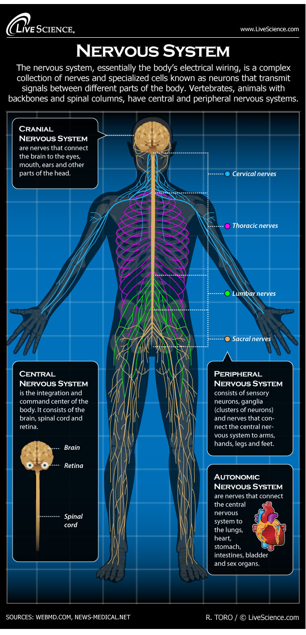
Human Nervous System - Diagram - How It Works | Live Science
Head anatomy: Muscles, glands, arteries and nerves | Kenhub The nerves in the head originate from three main sources: Trigeminal nerve (CN V) Facial nerve (CN VII) Cervical plexus The trigeminal nerve supplies sensory innervation to the forehead and cheek regions of the face via its ophthalmic and maxillary branches.
The nerves of the head and neck | Anatomy of the nerves of ...
Nerves and arteries of head and neck: Anatomy, branches | Kenhub

Anatomy of the Child's Nervous System

vagus nerve | Definition, Function, & Facts | Britannica
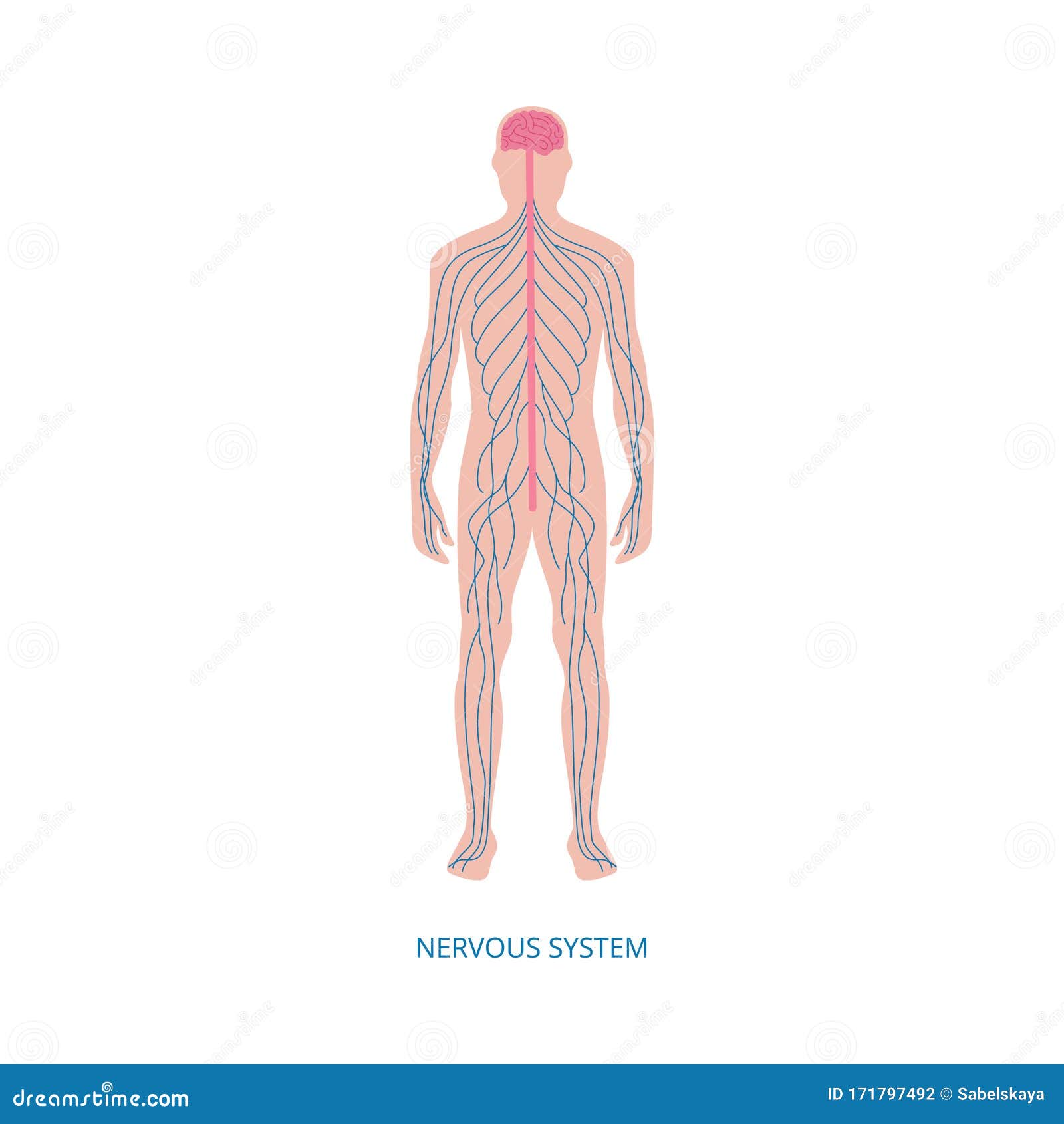
THE NERVOUS SYSTEM
15.2: Cranial Nerves - Biology LibreTexts

Trigeminal nerve diagram. ganglion, ophthalmic, mandibular ...
/GettyImages-87323698-5718ed505f9b58857db728ab.jpg)
Cranial Nerve Damage From Head Trauma
You've Got Some More Nerve(s): The Cranial Nerves
These Are the 12 Cranial Nerves and Their Functions
Superficial nerves of the face and scalp: Anatomy | Kenhub
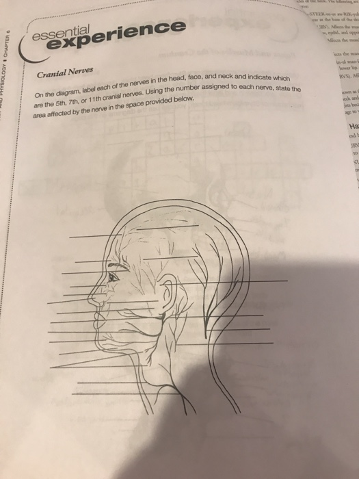
Solved On the diagram, label each of the nerves in the head ...
Nerves of the Head - TeachMeAnatomy
Cranial Nerve Anatomy / Cranial nerves | Iowa Head and Neck ...
/cranial-nerves-2-1e3d489c9104495dbcc609ea188af32d.jpg)
Names, Functions, and Locations of Cranial Nerves
Summary of the Cranial Nerves - TeachMeAnatomy
Central nervous system: Structure, function, and diseases
Cranial nerves: Anatomy, names, functions and mnemonics | Kenhub
Trigeminal Nerve: Function, Anatomy, and Diagram

Outline of a body showing the nervous system with ...
Cranial nerves quizzes and labeling exercises | Kenhub

Anatomy Of Nerves Of Body And Head Stock Illustration ...

Occipital Neuralgia
Nerves of the Head and Neck | Interactive Anatomy Guide
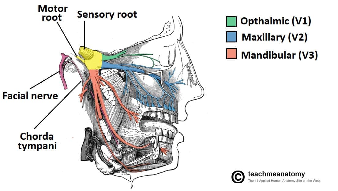
The Trigeminal Nerve (CN V) - Course - Divisions - TeachMeAnatomy

Head, Face, and Neck Nerves, labeling and function Diagram ...
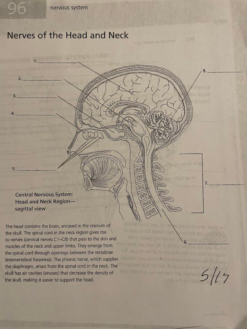
Solved 96 nervous system Nerves of the Head and Neck 8. 2 ...

Superficial nerves of the head and face Diagram | Quizlet

Cervical Plexus Anatomical Nerve Diagram Vector Illustration ...

What are the 12 cranial nerves? Functions and diagram

File:Head facial nerve branches.jpg - Wikimedia Commons
Cranial Nerve Anatomy / Cranial nerves | Iowa Head and Neck ...
0 Response to "45 nerves in the head diagram"
Post a Comment