43 in the diagram, where is the osteon?
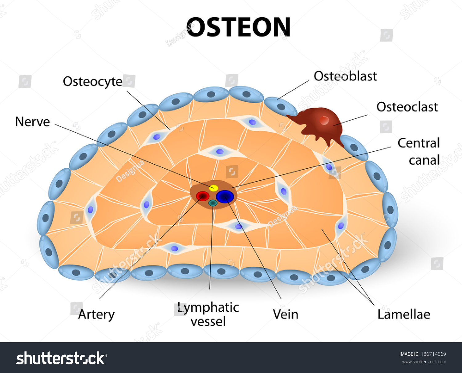
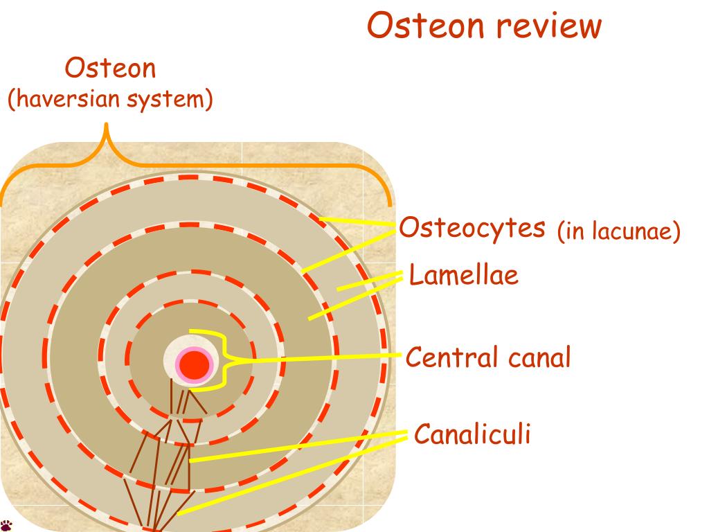
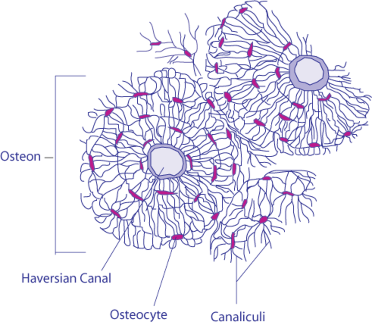
Osteons are cylindrical structures that contain a mineral matrix and living osteocytes connected by canaliculi, which transport blood. They are aligned parallel to the long axis of the bone. Each osteon consists of lamellae, which are layers of compact matrix that surround a central canal called the Haversian canal. -were the marrow is located Basic unit of structure in compact bone comprised of Lamellae, central canal and osteocytes. Osteocyte OSTEON BONE DIAGRAM: AN OSTEON BY ASHLEY HOLMES Central Canal -part of the a bone cell, forms when an osteoblast becomes embedded in the matrix it has secreted Lamellae layers on bone tissue found in compact bone
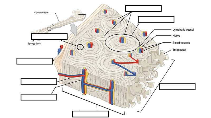
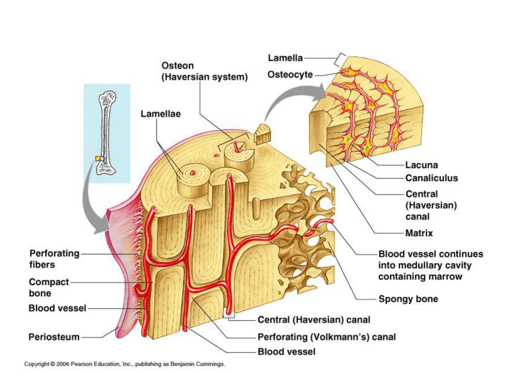
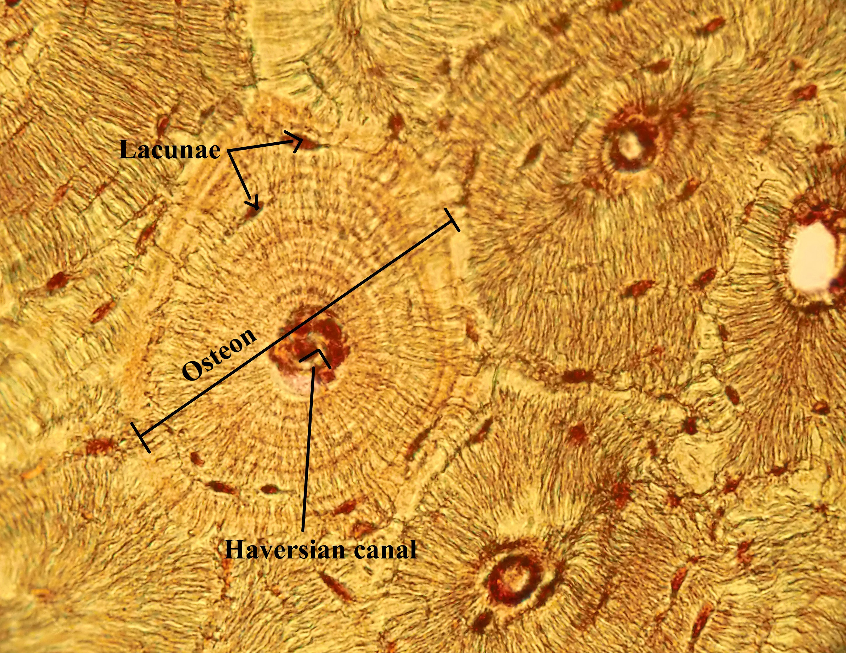
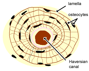
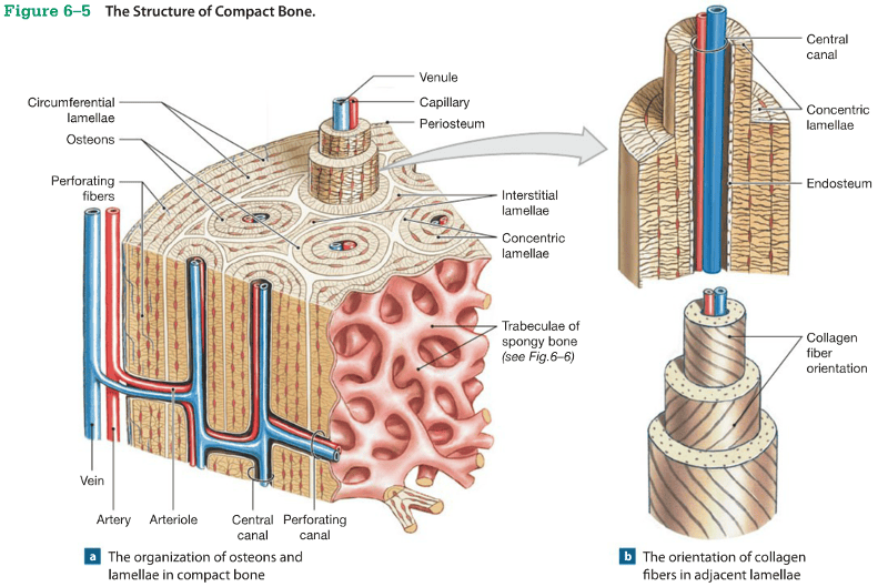
Compact bone consists of closely packed osteons or haversian systems. The osteon consists of a central canal called the osteonic (haversian) canal, which is surrounded by concentric rings (lamellae) of matrix. Between the rings of matrix, the bone cells (osteocytes) are located in spaces called lacunae.
In the diagram, where is the osteon?
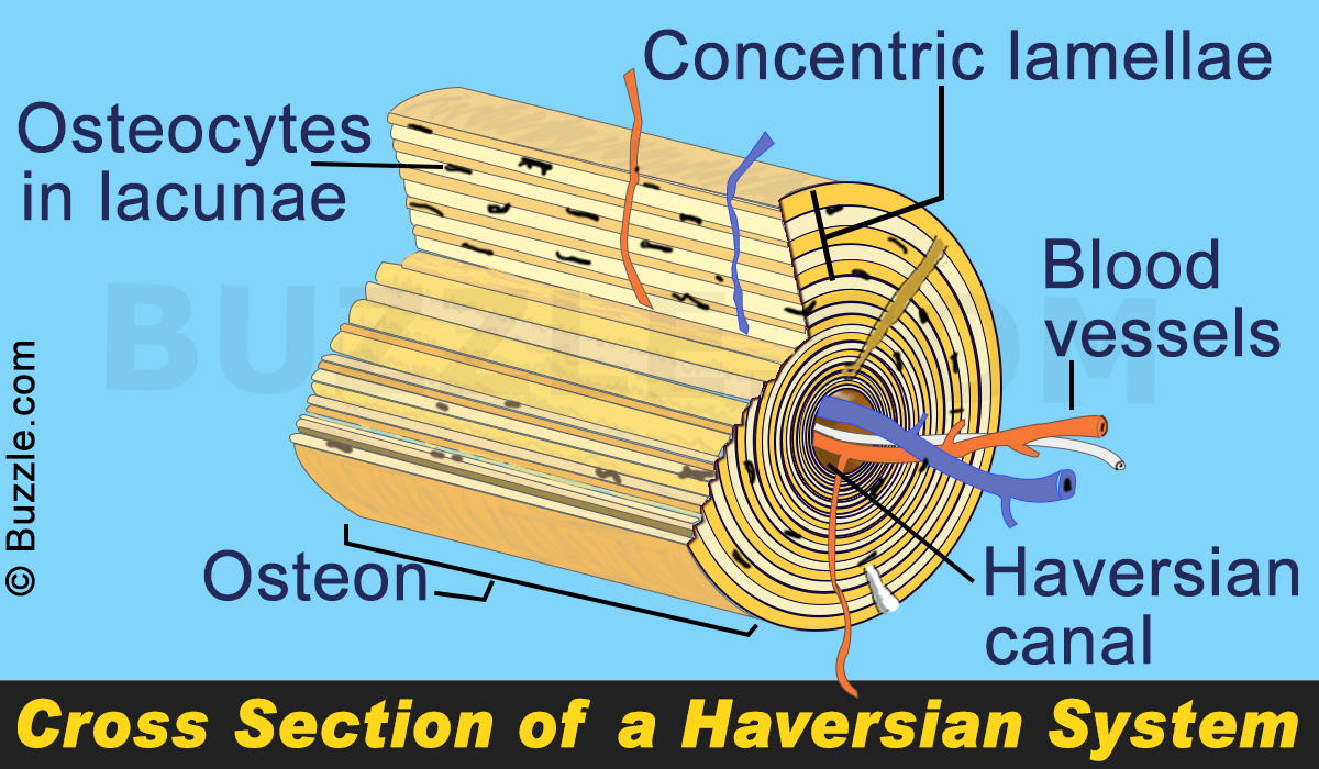
The structure including the central blood vessel and surrounding concentric bone tissue is called an osteon. What differentiates primary from secondary osteonal cortical bone is the way in which the osteon is formed and the resulting differences in the 2 nd level structure. It can be found under the periosteum and in the diaphyses of long bones, where it provides support and protection. Figure 6.12. Diagram of Compact Bone (a) This cross-sectional view of compact bone shows the basic structural unit, the osteon. (b) In this micrograph of the osteon, you can clearly see the concentric lamellae and central canals. Download scientific diagram | Schematic diagram of a forming osteon or bone remodelling unit. Osteoclasts (bone resorbing cells) cut a longitudinal tunnel through existing cortical bone. The ...
In the diagram, where is the osteon?. Osteon hard, dense bone tissue found on the outer surface of a bone bone found on the inside of bone; composed of lattice-like net… densely packed subunits of compact bone 5 Terms Vet217 PLUS Osteon Structure Lamellae Lacunae central canal (haversian canal) Concentric rings made up of groups of hollow tubes of bone mat… Compact bone is the denser, stronger of the two types of bone tissue ( (Figure) ). It can be found under the periosteum and in the diaphyses of long bones, where it provides support and protection. Diagram of Compact Bone. (a) This cross-sectional view of compact bone shows the basic structural unit, the osteon. Haversian system or osteon. This (Haversian system or osteon) is the structural unit of a compact bone matrix. They are the long cylindrical and branching structural unit that lies parallel to the long axis of the bone shaft. Each of the osteon or Haversian systems contains a centre canal or Haversian canal at the system's centre. The osteon or haversian system /həˈvɜːr.ʒən/ (named for Clopton Havers) is the fundamental functional unit of much compact bone. Osteons are roughly cylindrical structures that are typically between 0.25 mm and 0.35 mm in diameter.[1] Their length is often hard to define,[2] but estimates vary from several millimeters[3] to around 1 centimeter.[1]
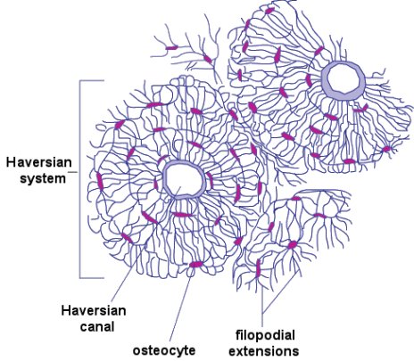
central part of the osteon. provides passageway for bones, nerve, and blood supply. surrounded by rings of Lamellae. Canaliculi. Microscopic canals within lamellae that link osteocyte. Sets found in the same folder. head/cranium bones. 17 terms. l_dewey1. The diagram above shows a longitudinal view of an osteon. Some, mostly older, compact bone is remodelled to form these Haversian systems (or osteons ). The osteocytes sit in their lacunae in concentric rings around a central Haversian canal (which runs longitudinally). It can be found under the periosteum and in the diaphyses of long bones, where it provides support and protection. Diagram of Compact Bone (a) This cross-sectional view of compact bone shows the basic structural unit, the osteon. (b) In this micrograph of the osteon, you can clearly see the concentric lamellae and central canals. LM × 40. osteon, the chief structural unit of compact (cortical) bone, consisting of concentric bone layers called lamellae, which surround a long hollow passageway, the Haversian canal (named for Clopton Havers, a 17th-century English physician).
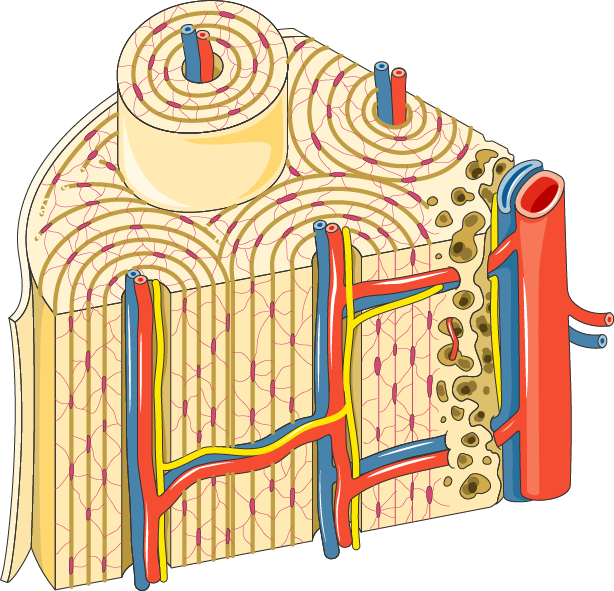
The osteons run parallel to the long axis of a bone. The long bones are composed of: Diaphysis Epiphysis Epiphyseal/growth plate Epiphyseal line Periosteum EndosteumWhile the diaphysis is the shaft that makes up the long axis of the bone, epiphysis refers to the ends of the bone. The ends are covered by a thin layer of compact bone. An osteon comprises a long, hollow central canal that is surrounded by concentric layers called lamallae. This central canal is referred to as the Haversian canal. The long axis of the osteon is parallel to the long axis of the bone. Each osteon has a cylindrical structure that consists of the following components: Download scientific diagram | 3: Diagram of an osteon, the primary structural unit of bone, with the concentric locations of osteocytes shown (Vaughan et al ... The diagram above shows a transverse view of an osteon ( Haversian system) - the basic unit of compact bone. diagram of haversian canal.An osteon comprises a long, hollow central canal that is surrounded by concentric layers called lamallae. This central canal is referred to as the Haversian canal. .
Osteon Development Structure Osteoblast Osteocyte ...
Question: - 3) C ( Osteon is a cylindrical structure present in the compact bone which contains H…View the full answer. Transcribed image text: } In the ...

Structure Of Compact Bone And The Osteon - sharedoc
UNIT 5/Chapter 5 - Review Guide - KEY. chapter_5_skeletal_system_review_guide_key.docx. File Size: 3324 kb. File Type: docx. Download File.

Anatomy Of Compact Bone. Compact Bone Tissue Osteon ...
The osteon consists of a central canal called the osteonic (haversian) canal, which is surrounded by concentric rings (lamellae) of matrix. People interested in compact bone diagram also searched for. The osteocytes are sitting in the lacunae and the canals are canaliculi, which interconnect the lacunae with the major vessels.

Compact Bone Diagram : The stability of a compact bone is ...
Lining the inside of the bone adjacent to the medullary cavity is a layer of bone cells called the endosteum (endo- = "inside"; osteo- = "bone"). These bone cells (described later) cause the bone to grow, repair, and remodel throughout life. On the outside of bones there is another layer of cells that grow, repair and remodel bone as well.
Bone Tissue (Guided Learning)
80% (5 ratings) Central canal is also called harversian canal.It is a circular canal …. View the full answer. Transcribed image text: entify the structures of an osteon Part A Drag the labels onto the diagram to identify the structures of an osteon. Reset Help central canal JOIN lacuna lamella canac Submit Request Answer.
Osteon Diagram
Osteon Definition another name for a Haversian system in bone Location Term Central canal Definition part of the osteon that is in the middle of the lamellae and contains blood vessels and nerves. These run longitudinally through the bone. Location osteocytes all bone cells belong to this group whether they build bone or destroy it. lacunae

Ostéon : définition et explications
Diagram of Compact Bone. (a) This cross-sectional view of compact bone shows the basic structural unit, the osteon. (b) In this micrograph of the osteon, you can clearly see the concentric lamellae and central canals.
Compact Bone Diagram Labeled / Labeled Diagram Of Femur ...
About Press Copyright Contact us Creators Advertise Developers Terms Privacy Policy & Safety How YouTube works Test new features Press Copyright Contact us Creators ...
5 Structure of the osteons of cortical bone (Junqueira and ...
53. Which of the labeled structures in the diagram are composed of trabeculae, which are bony structures that lack osteons? B ; F ·. Where in the figure is the ...
Osteon Diagram
Download scientific diagram | 3: Diagram of an osteon, the primary structural unit of bone, with the concentric locations of osteocytes shown (Vaughan et al.The microscopic structural unit of compact bone is called an osteon, or Haversian system. Each osteon is composed of concentric rings of calcified matrix called lamellae (singular = lamella).

Filippi, Jennifer / Useful Study Dox
Each osteon consists of concentric layers, or lamellae, of compact bone tissue that surround a central canal, the haversian canal. The haversian canal contains the bone's blood supplies. The boundary of an osteon is the cement line . Each haversian canal is surrounded by varying number (5-20) of concentrically arranged lamellae of bone matrix.

Compact Bone Diagram Unlabeled - Filippi, Jennifer ...
Each osteon is a compact cylinder of concentric lamellae. The only cells in an osteon are the osteocytes that are found on the edges of each lamella. Osteocytes are found in lacunae, which are the cell-shaped empty spaces that prevent the solid, mineralized extracellular material of bone from crushing the osteocytes.
PPT - Osteon development PowerPoint Presentation - ID:416574
Start studying Osteon Label. Learn vocabulary, terms, and more with flashcards, games, and other study tools.

Internal structure of a bone and an osteon
In the diagram, where is the osteon? B. In the diagram, where is the trabeculae? B. In the diagram, where is the zone of hypertrophic cartilage? A. In the diagram, this zone contains mostly dead chondrocytes surrounded by a calcified matrix. D. In the diagram, where is the zone of resting cartilage? A.

In The Diagram Where Is The Osteon - Atkinsjewelry
Download scientific diagram | Schematic diagram of a forming osteon or bone remodelling unit. Osteoclasts (bone resorbing cells) cut a longitudinal tunnel through existing cortical bone. The ...
Lab Activities - Human Anatomy Lab Manual
It can be found under the periosteum and in the diaphyses of long bones, where it provides support and protection. Figure 6.12. Diagram of Compact Bone (a) This cross-sectional view of compact bone shows the basic structural unit, the osteon. (b) In this micrograph of the osteon, you can clearly see the concentric lamellae and central canals.

Structure of Compact Bone Quiz
The structure including the central blood vessel and surrounding concentric bone tissue is called an osteon. What differentiates primary from secondary osteonal cortical bone is the way in which the osteon is formed and the resulting differences in the 2 nd level structure.

Haversian canal occurs in. toppr.com

In The Diagram Where Is The Osteon - Drivenheisenberg
Osteoblasts, Osteoclasts, Calcium, and Bone Remodeling ...
Cartilage, Bone & Ossification: The Histology Guide

osteon model | A&P.2.Skin.Bone.Muscle | Pinterest | Anatomy

osteon slide - YouTube

Osteon - Wikipedia

Where in the diagram is the endosteum located?
Soft Bone Tissue in a Triceratops Fossil - Proslogion

Pinned map of the United States of America

3: Diagram of an osteon, the primary structural unit of ...

Image result for osteon blank | Structure of bone, Red ...

Closeup of skeleton foot model

(A) Histological cross-section of cortical bone, showing ...

china drifter
In The Diagram Where Is The Osteon - Drivenheisenberg
osteon - Liberal Dictionary
Bone Doctor | Osteocytes | Louisville Orthopedic Surgeon
Osteon || Med-koM

Compact Bone Diagram - Compact Bone Structure Biology ...

Compact Bone Diagram Labeled - Osteon Wikipedia
Closeup of skeleton pelvic model

hello yellow on a small wall
BME 332: Bone Structure-Function

In The Diagram Where Is The Osteon - General Wiring Diagram
Chapter 6: Osseous Tissue and Bone Structure Flashcards ...
0 Response to "43 in the diagram, where is the osteon?"
Post a Comment