40 cell membrane diagram labeled
The parts of a plant cell include the cell wall, the cell membrane, the cytoskeleton. Leaves take in energy via sunlight and capture carbon dioxide from the air. Animal Cell Diagram Unlabeled — UNTPIKAPPS. Animal Cell Diagram Unlabeled — UNTPIKAPPS. pictures of plant and animal cells for kids to fill out …. Practice labeling the parts of the cell membrane Learn with flashcards, games, and more — for free.
Chapter 4: Membrane Structure and Function How are Cell Surfaces Specialized? Answer: Cell walls offer support and protection Cell Walls: • Found in bacteria, plants, fungi, & some protists • Composed of carbohydrates (e.g. cellulose, chitin), proteins, or inorganic molecules (e.g. silica) • Produced by the cell it protects/supports

Cell membrane diagram labeled
Fig. 2.3 Membrane structure. (A) The cell membrane showing in-tegral and peripheral proteins. (B) Intercalation of kinked unsaturated fatty acid chains and cholesterol molecules spaces out the phospho-lipid molecules and affects membrane fluidity. (C) Antibody molecules cross-linking peripheral proteins. This reduces the mobility of integral An additional non-living layer present outside the cell membrane in some cells that provides structure, protection, and filtering mechanism to the cell is the cell wall. Structure of Cell Wall. In a plant cell, the cell wall is made up of cellulose, hemicellulose, and proteins while in a fungal cell, it is composed of chitin. Drawing Diagrams Diagram 1: Human cheek cells The photo below is of a cluster of human cheek cells that have been stained and magnified 400 times. On your lab sheet draw the cluster of cells and label one cell with the following structures: cell membrane - the cell membrane is the outer edge of the cell
Cell membrane diagram labeled. Membrane Structure and Function All cells have a plasma or cell membrane , which contains the cell. Scanning electron micrograph (SEM) of adipocytes (Ad) Membrane Structure and Function Prokaryotic Cells: Bacteria. The Formation of Cell Membranes is Crucial to Life. May 18, 2013 · Talking related with Cell Membrane Labeling Worksheet, we've collected some similar images to complete your references. cell membrane diagram labeled, cell organelle labeling worksheet and animal cell coloring answers are three main things we want to present to you based on the post title. Match the cell membrane structure or its function with the correct letter from the cell membrane diagram. Letter Structure/Function ... Color and label the cell in an isotonic environment light blue, the hypotonic environment yellow, and the hypertonic environment light green. When drawing a diagram of a ... Our current model of the cell membrane is called the Singer-Nicholson fluid mosaic model ... Lines touch the labeled.31 pages
Here are a number of highest rated Simple Cell Membrane Labeled pictures upon internet. We identified it from obedient source. Its submitted by government in the best field. We believe this nice of Simple Cell Membrane Labeled graphic could possibly be the most trending topic taking into consideration we part it in google lead or facebook. We are pleased to provide you with the picture named Cell Membrane Diagram.We hope this picture Cell Membrane Diagram can help you study and research. for more anatomy content please follow us and visit our website: www.anatomynote.com. Anatomynote.com found Cell Membrane Diagram from plenty of anatomical pictures on the internet.We think this is the most useful anatomy picture that you need. The cell membrane, also called the plasma membrane, is a thin layer that surrounds the cytoplasm of all prokaryotic and eukaryotic cells, including plant and animal cells. It is a selectively permeable cell organelle,allowing certain substances inside the cell while preventing others to pass through and thus is analogous to a barrier or gatekeeper in their function. Start studying Labeling a cell membrane. Learn vocabulary, terms, and more with flashcards, games, and other study tools.
This diagram shows the structure of a phospholipid. The hydrophilic head group is shown as. Figure 1. Phospholipid Structure. A phospholipid molecule consists ... Thin layer of protein and fat that surrounds the cell is the cell membrane. More information Printable labeled and unlabeled animal cell diagrams, with list of parts and definitions Cell Membrane Detailed Diagram Labeled. B) Tumor cells carrying the BCN group via natural glycometabolic labeling by. Unlabeled Animal Cell - Cliparts.co 3 Match the cell membrane structure or its function with the correct letter from the cell membrane diagram. Letter Structure/Function Letter Structure/Function G Attracts water F Repels water I Helps maintain flexibility of membrane G & F Make up the bilayer C & E Involved in cell-to-cell recognition One of the most important considerations for the design of the logo is the color palette. Help texture speaking through design elements with transparency. When you are designing a logo, consider whether or not a generic or unique design. Cell Membrane Diagram Labeled via Cell Membrane Coloring Worksheet Answers via
Corona Contact detection app on Android
The cell membrane plasma membrane is a thin semi permeable membrane that surrounds the cytoplasm of a cell. Animal and plant cells worksheet. Label 1 is label 2 is. The fundamental structure of the membrane is the phospholipid bilayer which forms a stable barrier between two aqueous compartments. Thin membranes bound all living cells and many ...

using mobile phone photo by screen post
When salt is poured on a slug, it dies because. salt moves into the slug, poisoning it. water moves into the slug, causing it to swell. water moves out of the slug, causing dehydration. salt moves out of the slug, depriving it of essential minerals. Movement of the small molecules from left to right across the membrane. requires active transport.

Amazon Go
Cell membrane drawing labeled. A labeled diagram of the animal cell and its organelles …. 4.2.1 cell membrane (plasma membrane) each cell has a limiting boundary, the cell membrane, plasma membrane or plasmalemma. The cell membrane is a multifaceted membrane that envelopes a cell's cytoplasm.

Cell Membrane
Feb 06, 2017 · Diagram of the human cell illustrating the different parts of the cell. Cell Membrane The cell membrane is the outer coating of the cell and contains the cytoplasm, substances within it and the organelle. It is a double-layered membrane composed of proteins and lipids. The lipid

Note 9
Figure: Labeled diagram of plant cell, created with biorender.com The typical characteristics that define the plant cell include cellulose, hemicellulose and pectin, plastids which play a major role in photosynthesis and storage of starch, large vacuoles responsible for regulating the cell turgor pressure.

Motorola V3
In other words, a diagram of the membrane (like the one below) is just a snapshot of a dynamic process in which phospholipids and proteins are continually ...

Discovery and Structure of Cells | Biology | Visionlearning
Feb 01, 2021 · As observed in the labeled animal cell diagram, the cell membrane forms the confining factor of the cell, that is it envelopes the cell. An additional non-living layer present outside the cell membrane in some cells that provides structure, protection, and filtering mechanism to the cell is the cell wall.

3D Model of Animal Cell - YouTube
Full-Length Text • Here, we'll learn about the cell membrane, which separates the inside of the cell from its external environment. • First, start a table to summarize the major features of the plasma membrane. • Denote that the plasma membrane comprises: - Phospholipids, which have hydrophilic heads and hydrophobic tails.
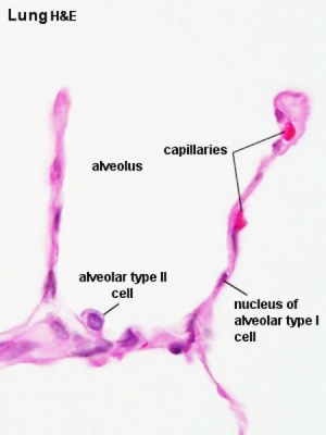
File:Alveolar-sac-01.jpg - Embryology
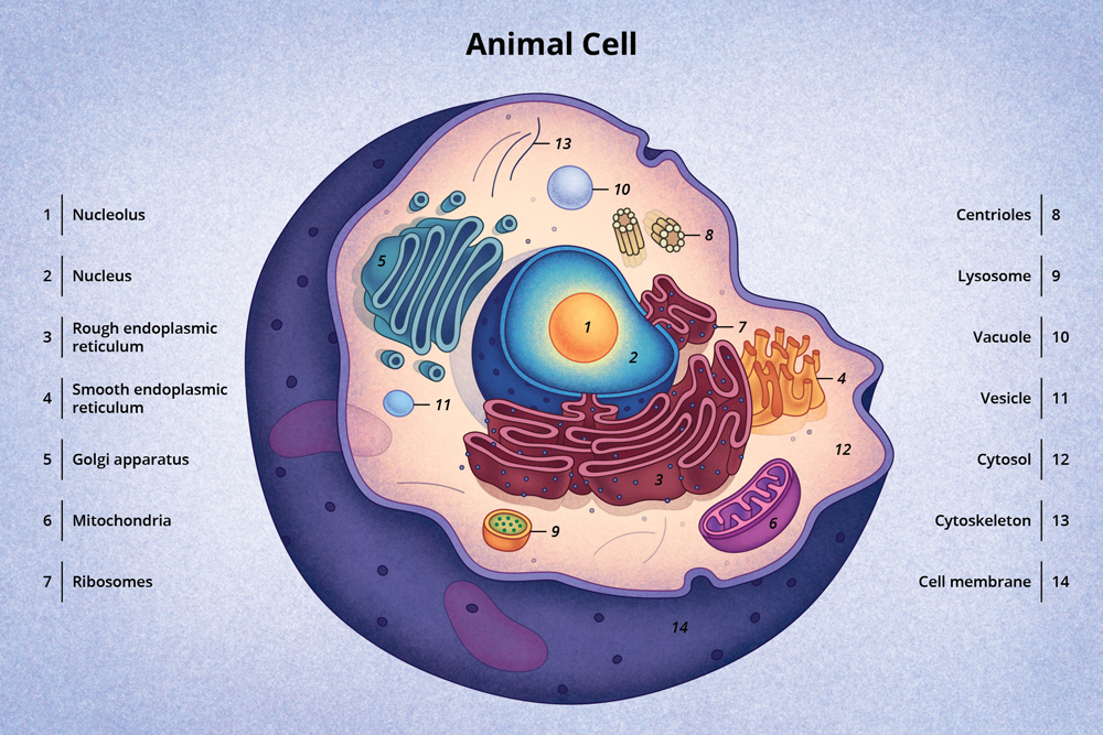
Animal cells are eukaryotic cells. They contain membrane-bound nuclei. Diagram of Animal Cell is beneficial in understanding the structure and functions of an animal. This article comprehends a brief explanation of the different parts of an animal cell with a well-labelled diagram.

Twitter is a good platform and a micro social media for trending news and current affairs.
This cell membrane provides a protective barrier around the cell and regulates which materials can pass in or out. Structure and Composition of the Cell Membrane. The cell membrane is an extremely pliable structure composed primarily of two layers of phospholipids (a "bilayer"). Cholesterol and various proteins are also embedded within the ...
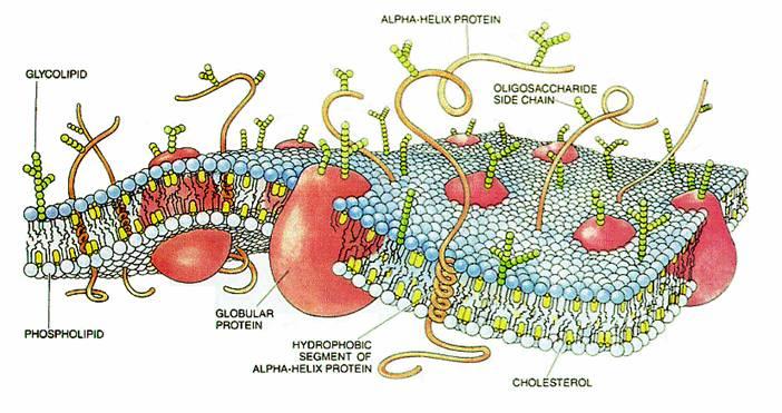
Diagram of a cell membrane with labels | NIST
Jul 13, 2020 - This Pin was discovered by Michelle Figueroa. Discover (and save!) your own Pins on Pinterest.

September 2020 planner and techy tools tools
Anatomynote.com found Simple Diffusion Across The Cell (plasma) Membrane Diagram from plenty of anatomical pictures on the internet. We think this is the most useful anatomy picture that you need. You can click the image to magnify if you cannot see clearly. This image added by admin. Thank you for visit anatomynote.com.
Discovery and Structure of Cells | Biology | Visionlearning
Fluid mosaic model: cell membranes article. It may seem like the human body is made up of a chaotic mix of random parts, but that's not the case. The liquid nutrients, cell machinery, and blueprint information that make up the human body are tucked away inside individual cells, surrounded by a double layer of lipids.
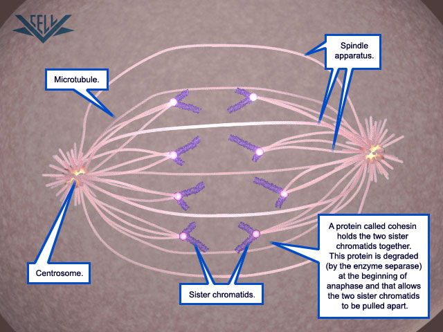
VCAC: Cellular Processes: Mitosis: Advanced Look: Mitosis
Cell Membrane Detailed Diagram Labeled. A diagram of the plasma membrane of a cell. The phospholipid bilayer separates the cytoplasm (below) from the extracellular fluid (above). This image is created by Wikimedia Commons user LadyofHats Mariana Ruiz, who released it into the public domain.

Taking a Photo with a Google Android Smartphone
Cell membrane, thin membrane that surrounds every living cell. The cell membrane functions as a barrier, keeping cell constituents in and unwanted ...
Print Structure and Function of Plants (Lab Practical #1 ...
cell membrane organelles cytoplasm . CELL STRUCTURE AND FUNCTION CHART PLANT CELL ANIMAL CELL . 1. Cell Wall •(Plants only) inflexible barrier “protecting” the cell and giving it support. Is not selectively permeable. It is a rigid structure.

Corona contact detection app
The cell membrane is a multifaceted membrane that envelopes a cell's cytoplasm. It protects the integrity of the cell along with supporting the cell and helping to maintain the cell's shape. Proteins and lipids are the major components of the cell membrane. The exact mix or ratio of proteins and lipids can vary depending on the function of a ...
0 Response to "40 cell membrane diagram labeled"
Post a Comment