42 spinal nerve root diagram
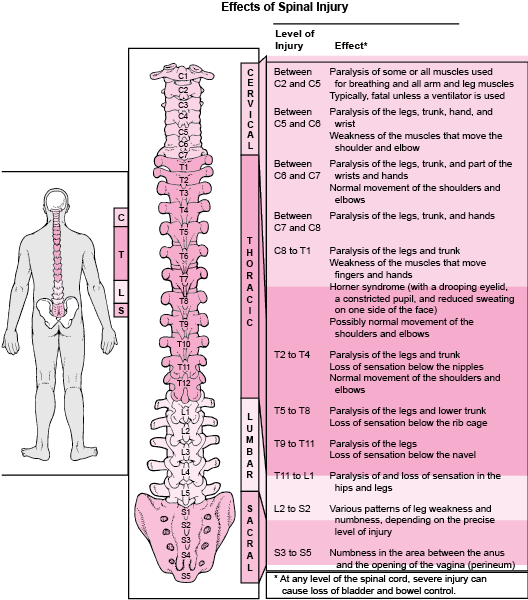
The spinal cord can be equally divided along the midline dorsoventral axis by drawing a line through the depression known as the dorsal and ventral median sulci. On each half of the spinal cord, a ventrolateral and dorsolateral sulcus is appreciated at the sites from which the ventral and dorsal nerve roots leave Root of the neck. posterior: T1 spinal nerve; lateral: suprapleural membrane, vertebral artery; anterior: carotid sheath, stellate ganglion is located opposite to the neck of the 1st rib, phrenic nerve, scalenus anterior; Thorax. posterior: intercostal nerves, intercostal vessels,
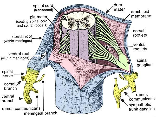
Thus there are 31 pairs of spinal segments. The dorsal nerve roots enter the spinal cord along the postero-lateral sulcus and ventral roots along the anterolateral sulcus. The dorsal root shows a swelling called dorsal nerve root ganglion (spinal ganglion). This ganglion contains cell bodies of pseudounipolar sensory neurons.
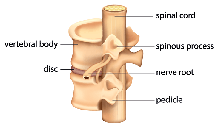
Spinal nerve root diagram
Spinal nerves are mixed nerves that transmit motor, sensory, and autonomic signals between the central nervous system and the periphery Each spinal nerve carries afferent (sensory) fibers and efferent (motor) fibers to and from the spinal cord, the former of which comprise the posterior/dorsal rootsEach posterior root presents a ganglion as it emerges from the intervertebral foramen. They form from nerve roots that branch from your spinal cord. Spinal nerves are named and grouped by the region of the spine that they're associated with.Spine and sensation · Dermatomes list The rootlets unite to form an anterior (ventral) or posterior (dorsal) root of a spinal nerve. The anterior/ventral root contains efferent nerve fibres, which carry stimuli away from the CNS towards their target structures.The cell bodies of the anterior root neurons are located in the central grey matter of the spinal cord.
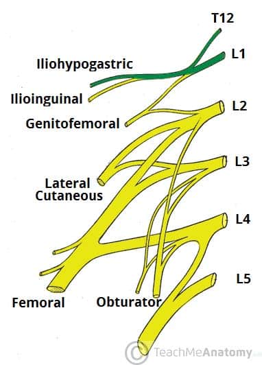
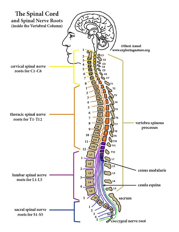
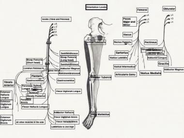
Spinal nerve root diagram. The relevant anatomy of the spinal nerve-muscular innervation of the back is centered around the lumbar spinal nerves, peripheral nerves of the lumbar plexus, spinal cord, and lumbar vertebral column. Within the lumbar region, the vertebral bodies are larger than in the thoracic and cervical regions due to the lumbar spine being designed for weight-bearing purposes. Course and branches of thoracic spinal nerve: This diagram depicts the ... A myotome is the group of muscles that a single spinal nerve root innervates. A dorsal root ganglion is the one associated with the dorsal or posterior root of the nerves originating from the spinal cord. All the posterior roots of spinal nerves contain a ganglion. As the dorsal or posterior root of a spinal nerve is primarily sensory, the dorsal root ganglion contains cell bodies of these sensory nerve fibers. The lumbar nerve roots exit beneath the corresponding vertebral pedicle through the ... Simplified coronal diagram of lumbosacral plexus, depicted on a ...
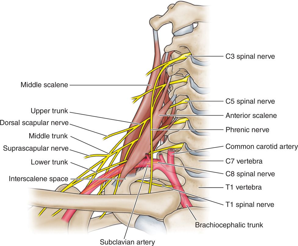
The long thoracic nerve is a lateral branch of the brachial plexus, which arises from the anterior rami of spinal nerves C5, C6 and C7. These nerve roots commence deep to the scalenus medius muscle to form the trunk of the long thoracic nerve Due to its relatively superficial location, almost the entire course of the nerve is easily visible in cadavers. Projections of the spinal cord into the nerves (red motor, blue sensory). Schematic diagram of cervical plexus. Dissection images. The thoracic part of the spinal cord gives rise to twelve pairs of thoracic spinal nerves. The anterior/ventral rami of the first eleven thoracic spinal nerves give rise to eleven intercostal nerves.The twelfth nerve is located inferior to the last rib and is thus called the subcostal nerve.. Upon arising, each intercostal nerve is connected to its corresponding sympathetic ganglion (of the ... The L4 spinal nerve roots exit the spinal cord with the help of small bony opening on the right and the left side of our spinal canal. Must Read Best Ayurveda Treatment For Sciatica Pain. The areas of our skin that receive sensations through L4 spinal nerve is known as L4 dermatome.
Traditionally the accessory nerve has been described as having both spinal and cranial roots. Consequently, the nerve is commonly discussed according to the two divisions (i.e. cranial and spinal roots). The cranial division (internal ramus) emerges from the medulla oblongata of the brainstem at the level of the nucleus ambiguus. A myotome is the group of muscles on one side of the body that are innervated by one spinal nerve root. During a physical exam, your healthcare provider would consider the location of myotomes and dermatomes to identify the specific spinal nerve (s) that may underlie problems such as muscle weakness and sensory changes. 2. A complete review of spinal anatomy and back pain, including the spinal cord and spinal nerve roots, with a look at herniated discs and pinched nerves. Spinal meninges (diagram) The spinal cord and spinal nerve roots are wrapped within three layers called meninges. The outermost is the dura mater, underneath it is the arachnoid mater, and the deepest is the pia mater. Dura mater has two layers (periosteal and meningeal), between which is the epidural space.
Motor nerve fibers, like those found in the brachial plexus, arise from cells within the basal plate of the developing spinal cord and emerge to the ventral nerve root. The sensory nerve fibers found in the dorsal nerve root originate from neural crest cells. The dorsal nerve root will grow toward the ventral nerve root and will eventually join ...
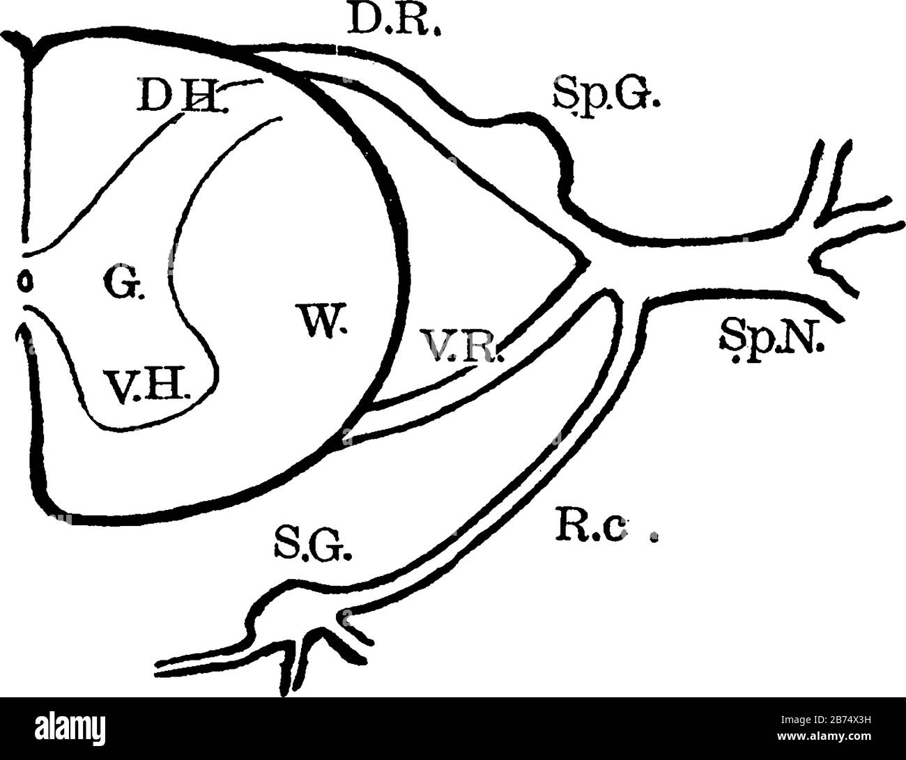
Diagram Showing Anatomy Of The Spinal Nerve Roots And Adjacent Parts Vintage Line Drawing Or Engraving Illustration Stock Vector Image Art Alamy
The height to which the agent ascends is controlled by the amount injected and the position of the patient. Sensation is lost inferior to the epidural block. Anesthetic and analgesic agents can also be injected through the posterior sacral foramina into the sacral canal around the spinal nerve roots ( transsacral epidural anesthesia) . Epidural ...
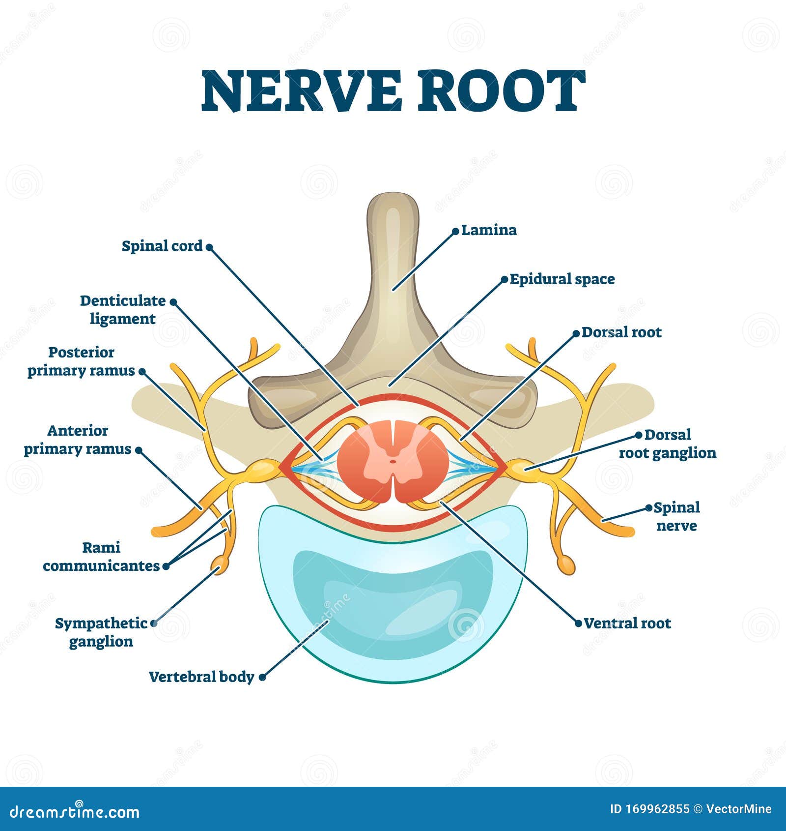
Nerve Root Anatomical Structure Labeled Cross Section Stock Vector Illustration Of Back Design 169962855
The 10 spinal laminae of the spinal cord are shown on a second diagram about the grey matter of the spinal cord. ... two axial sections of the spinal cord and adjacent structures allow the organisation of a spinal nerve to be displayed with its various branches (sensitive posterior root, anterior motor root, meningeal branch, muscular branches ...
The pudendal nerve originates from the sacral spinal nerves and innervates the descending colon, rectum, urinary bladder, and genitals. Coccygeal Spinal Nerves. There is 1 coccygeal spinal nerve pair.
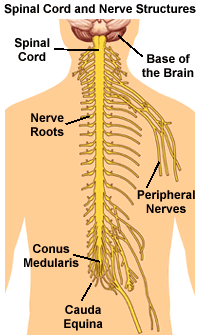
Spinal nerve, in vertebrates, any one of many paired peripheral nerves that arise from ... Near the spinal cord each spinal nerve branches into two roots.
The cervical nerves arise from the spinal cord in the form of rootlets, or fila radicularia, smaller neuron bundles that coalesce to form roots. For each spinal nerve, an anterior and posterior root join to form the completed nerve. Shortly after branching out of the spinal cord, the cervical nerves form the cervical and brachial plexuses.
Aug 18, 2012 — image to the left, the nerve roots (in red) branch off from the spinal cord to create the electrical wires of the PNS.
Each spinal nerve root has a corresponding medullary artery. A vasocorona surrounding the conus medullaris and the high degree of arterial anastomoses among the nerve roots predispose the vasculature patterns to significant diversity. Lymphatic capillaries occur nearly everywhere in the body except for a small number of sites, including the ...
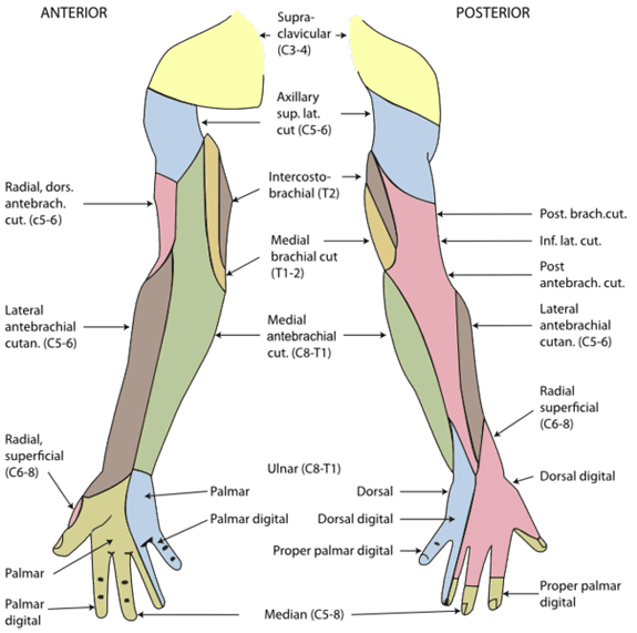
Each spinal nerve except C1 receives sensory input from a specific area of skin called a dermatome.22 A dermatome map (fig. 13.19) is a diagram of the cutaneous regions innervated by each spinal nerve. Such a map is oversimplified, however, because the dermatomes overlap at their edges by as much as 50%.

Anatomical Chart Spinal Nerves Spinal Nerve Root Innervation Chart Transparent Png 720x540 Free Download On Nicepng
The rootlets unite to form an anterior (ventral) or posterior (dorsal) root of a spinal nerve. The anterior/ventral root contains efferent nerve fibres, which carry stimuli away from the CNS towards their target structures.The cell bodies of the anterior root neurons are located in the central grey matter of the spinal cord.
They form from nerve roots that branch from your spinal cord. Spinal nerves are named and grouped by the region of the spine that they're associated with.Spine and sensation · Dermatomes list

Orthobullets There Are 2 Key Differences Between The Cervical And Lumbar Spine With Respect To Pathology And Level Affected 1 Pedicle Nerve Root Mismatch In The Cervical Spine C6 Nerve Root Travels
Spinal nerves are mixed nerves that transmit motor, sensory, and autonomic signals between the central nervous system and the periphery Each spinal nerve carries afferent (sensory) fibers and efferent (motor) fibers to and from the spinal cord, the former of which comprise the posterior/dorsal rootsEach posterior root presents a ganglion as it emerges from the intervertebral foramen.

Nerve Root Anatomical Structure Labeled Cross Section Stock Vector Illustration Of Back Design 169962855

Archive Image From Page 628 Of Cunningham S Text Book Of Anatomy 1914 Cunningham S Text Book Of Anatomy Cunninghamstextb00cunn Year 1914 Entering Posterior Nerve Root Amt Rooi Fig 528 Diagram Of The Spinal Origin Of The

/spinal-column--illustration-487736937-5a6e4ceceb97de0037ea5bb5.jpg)
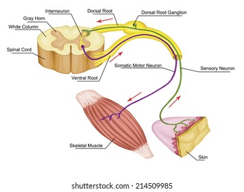




:background_color(FFFFFF):format(jpeg)/images/library/11473/spinal-membranes-and-nerve-roots_english.jpg)
:background_color(FFFFFF):format(jpeg)/images/article/en/spinal-nerves/91FC7O6eThfbi3VhQv2fMw_RbZ9HRprGLCiFN80S7sqjw_Posterior_cutaneous_branch_of_intercostal_nerve_02.png)






0 Response to "42 spinal nerve root diagram"
Post a Comment