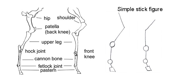
42 horse leg anatomy diagram
Of all the horse's biomechanical systems, perhaps one that is least understood or looked at is the lymphatic system. Much of our current research on equine lymphology has been conducted at the Veterinary University at Hannover in Germany, where they have discovered significant differences between the equine lymphatic system and that of the ... Buy Basic Anatomy Physiology Book Online At Low Prices In India Basic Anatomy Physiology Reviews Ratings Amazon In from images-na.ssl-images-amazon.com Explore resources and articles related to the human body's shape and form, including organs, skeleton, muscles, blood vessels, and more. Read up on the ankle and how it works.
Specific terms and specialized language are used to describe equine anatomy, different life stages, and colors and breeds.. Lifespan and life stages. Depending on breed, management and environment, the modern domestic horse has a life expectancy of 25 to 30 years. Uncommonly, a few animals live into their 40s and, occasionally, beyond. The oldest verifiable record was "Old Billy", a 19th ...

Horse leg anatomy diagram
What do you know about horses? Would you be able to pass this quiz on general equine anatomy? As it pertains to this quiz, you will be required to know the body part of the horse on the top line where the neck ends, and the back begins, what the middle joint of the back leg is called, and what is the highest point of the rump. You indeed have to take this sensational quiz. Horse Body Parts · Equine Forelimb Anatomy · Horse Hoof Anatomy · Horse Skeleton · Horse Teeth Anatomy · Great Anatomy Reference Books ... The limbs of the horse are structures made of dozens of bones, joints, muscles, tendons, and ligaments that support the weight of the equine body.
Horse leg anatomy diagram. The bone at the top of the leg. An atlas of cat anatomy. The lower extremity, commonly referred to as the leg, contains four bones (the femur, the patella, the tibia, and the fibula) and bends at the hip, the knee, and the ankle. This diagram of a feline skeleton shows you where all of your cat's bones are. Horse Diagrams - The Hoof. This is a detailed diagram of a horse's hoof. This will help me apply this to my integument presentation. We'll break down the anatomy and function of the upper leg, knee, lower leg, ankle, and foot. Inflammation in a tendon is known as tendinitis a tendon is a specialized structure that attaches mus. Casaraguru / istock it wasn't that long ago that if a horse broke a leg, euthanasia was the only. Some redness and tumefaction may have em. Muscle anatomy reference charts Author: Molly Smith DipCNM, mBANT • Reviewer: Dimitrios Mytilinaios MD, PhD Last reviewed: November 03, 2021 Reading time: 3 minutes If you've ever attempted to learn the origins, insertions, innervations, and functions of all 600+ muscles in the body… you'll know what a soul-destroying task it can be.
Horse Anatomy, Leg Anatomy, Animal Anatomy, Human Anatomy, Horse Information, Horse. Horse Anatomy Pictures-Think Like a Horse-Rick Gore Horsemanship ®. Horse Leg Bone Diagram - Forever Horses Anatomy Of The Equine Hindleg Horse Anatomy Large Animal Vet Equine Vet Tech : Thanks a lot for this tutorial, the hardest part for me was the legs on drawing horses! Search from 685 Horse Anatomy stock photos, pictures and royalty-free images from iStock. ... Vintage engraving of a Diagram of a horse's leg and foot. This is most commonly observed at night time, when it's dark and the temperature is cooler. This is the deeper REM sleep. The horse's muscles relax and brain waves shift in order to give the horse a complete rest. This sleep can last for about 2 to 3 hours, or as short as a few minutes, depending on the horse and its surroundings.
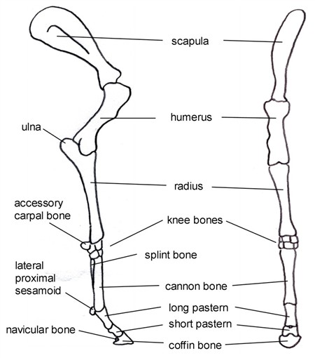
Important parts of the horse's forelimbs. The horse does not have a collarbone, so the front legs are not attached by joints, but rather to a ... The muscles of the lower limb are numerous and complex. Their origins and insertions are difficult to remember, and they are best considered as parts of general functional groups. iliopsoas psoas major psoas minor iliacus buttocks gluteal r... The lower leg makes up a large portion of an individual's overall body weight. It is an essential structure for any weight-bearing activity, such as walking, stand, running, or jumping. Common conditions that affect the lower leg include stress fractures , compartment syndrome , shin splints , and muscle tears. Develop an understanding of the causes of equine lameness and methods of treatment. Page 3. Parts of the Horse. Page 4 ...
Equine Tarsal Region Anatomy A Three Dimensional Volumetric Download Scientific Diagram
Skeletal system. This content is taken from our book, Managing Pig Health, the industry leading pig publication. Available now from 5mBooks.com. The structure of a bone and joint are shown in Fig.1-9. A joint consists of the ends of two bones held together by ligaments and muscles, surrounded by a strong membrane and covered with smooth ...
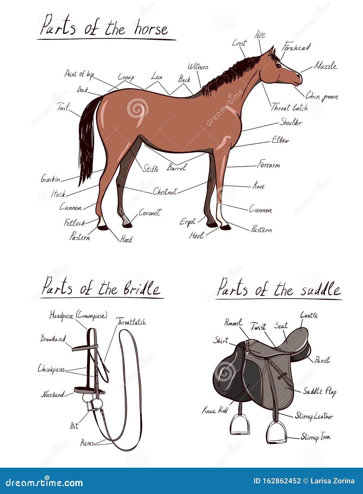
Parts Of Horse Saddle Bridle Set Equine Anatomy Equestrian Scheme Text Stock Illustration Illustration Of Riding Ranch 162862452
Equine anatomy refers to the gross and microscopic anatomy of horses, ponies and other equids, including donkeys, mules and zebras. While all anatomical ...

Vitals Anatomy Horse Side Vet Guide Horse Anatomy Horse Health Horse Facts
Anatomical diagram showing a front view of muscles in the human body. In the back and… Baca selengkapnya Basic Muscles In The Body Diagram / Horse Leg Anatomy - Front and Rear Leg Anatomy : *the origin, insertion, and belly.
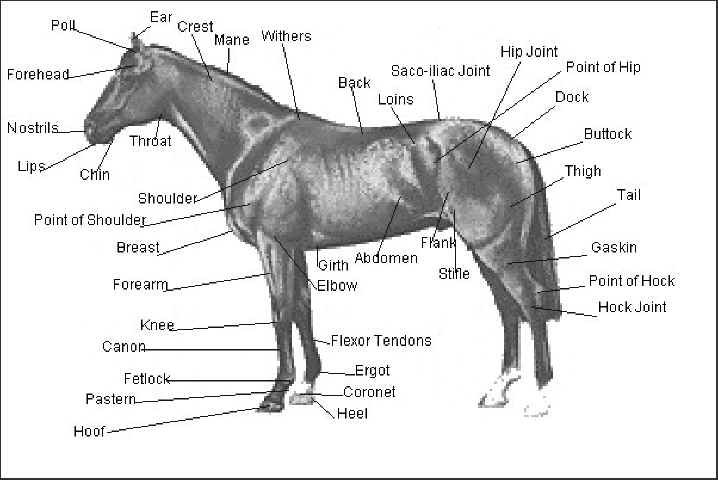
External Equine Anatomy All Things Equine
Anterior and Posterior Examples Anterior. The sternum, or breastbone, is anterior to the vertebral column.Additionally, concerning the rest of the body, the great saphenous vein (GSV) of the leg ...
Parts Of A Horse A Complete Guide With 3d Visible Horse
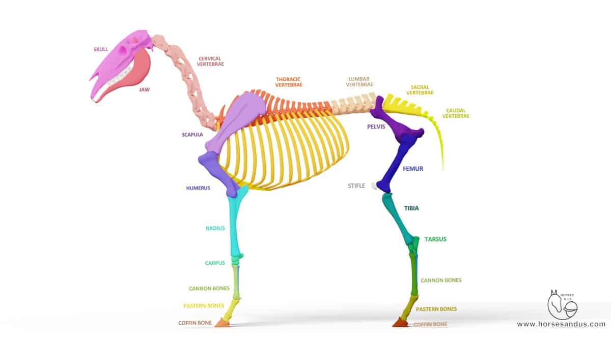
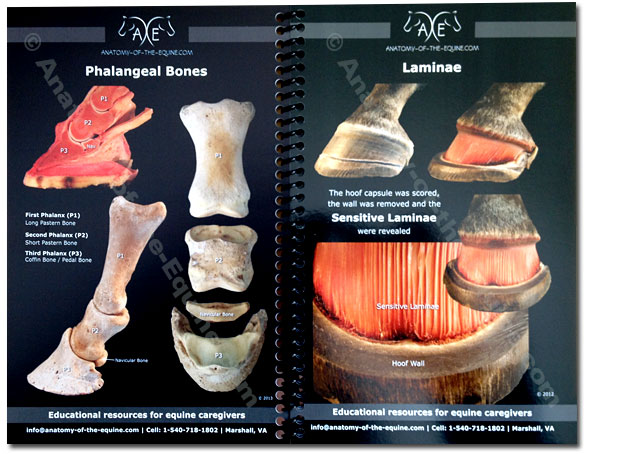
Equine anatomy - Illustrated atlas of the bones of the horse. This module of vet-Anatomy presents 135 labeled anatomical illustrations of the osteology of the horse, specially illustrated and selected for veterinary students and equine veterinarians.
How To Draw Horses Legs The Easy Way
A horse's knee is several bones held together by small muscles, tendons, and ligaments. The bones in the knee are similar to the bones of a human's wrists. The stifle joint in the back leg is actually closer in structure to a human knee.
2
Femur Anatomy: Overview. Bones come in a variety of shapes and sizes that allow them to function in different ways. Some, like the bones in our fingers, are small to allow for dexterous movements ...
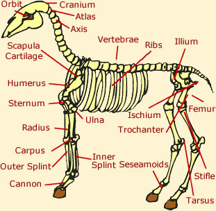
Horse Anatomy Skeleton Anatomy Diagram Of A Horse
The ischioanal fossa occupies most of the anal triangle. In a transverse section through the pelvis (in a lithotomy position), it has a horse shoe appearance, while in a coronal section (through a vertically erect individual) each fossa appears roughly pyramidal. The ischioanal fossa is limitted and comprised of the following structures:

Horse Anatomy Images Stock Photos Vectors Shutterstock
Normal centaur anatomy is a horse body with a humanoid torso & head that replaces the horse neck & head. Physiologically it works like a horse because the main body and legs are arranged like a horse. It's bones and muscles are those of a horse, so it can run like a horse. Human anatomy & physiology is uniquely adapted to an upright posture and ...
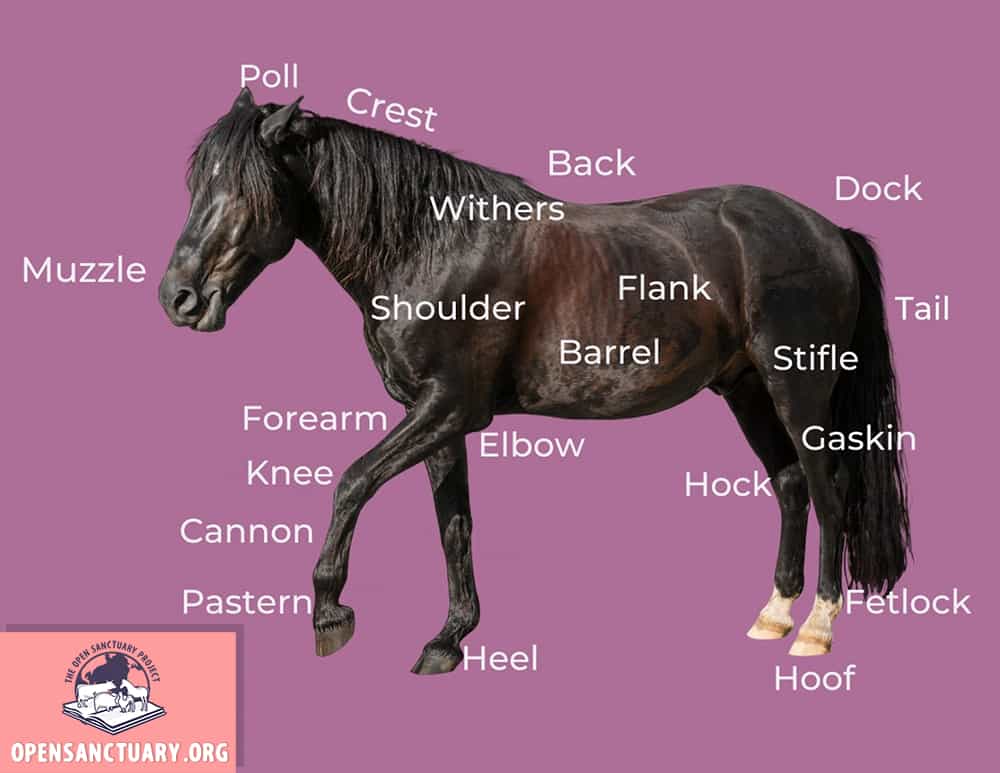
Basic Horse Anatomy Part 1 The Open Sanctuary Project
The hoof is the foot of the horse, consisting of a hard exterior and softer interior. Similar to a fingernail but much stronger. Knee. The knee (carpus) is a large, bending joint of the front leg. It functions more like a wrist than a knee. Muzzle. The muzzle consists of the nose, mouth, and chin of a horse. Pastern.

Horse Leg Bone Anatomy Back View
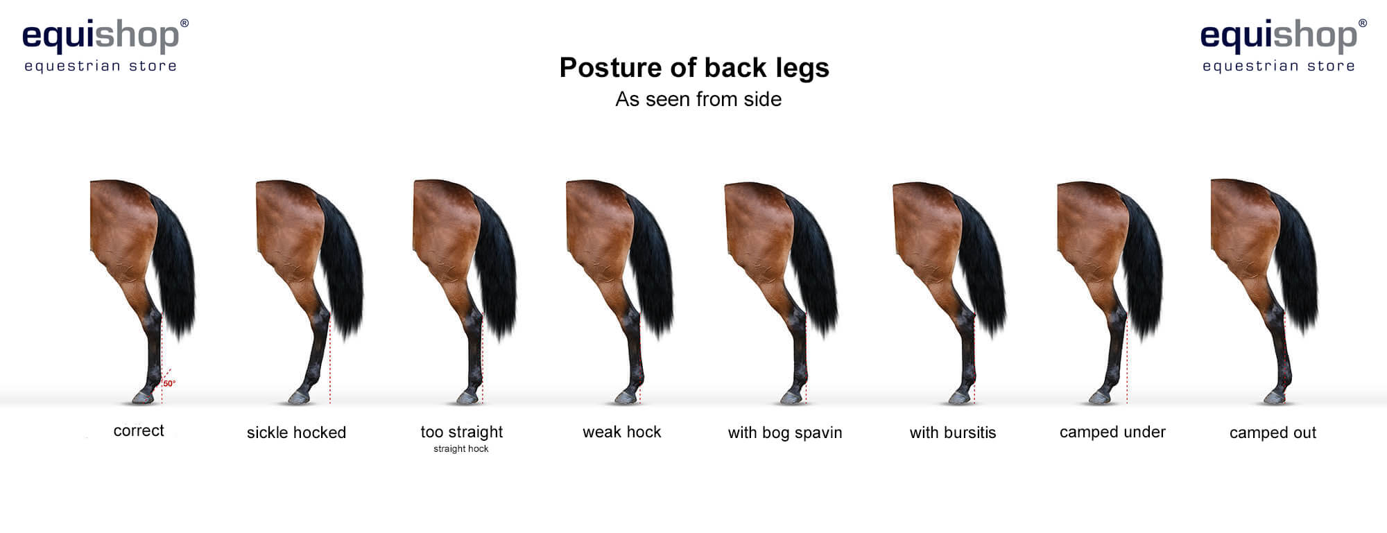
This is why it's so crucial for the horse's back legs to be as under the barrel as possible – this is how the "table" shortens and your body ...

Equine Tarsal Region Anatomy A Three Dimensional Volumetric Download Scientific Diagram
Bovine anatomy - Illustrated atlas. This module of vet-Anatomy provides the basics on the anatomy of the bull for students of veterinary medicine. This veterinary anatomical atlas includes 27 scientific illustrations with a selection of labelled structures to understand and discover animal anatomy (skeleton, bones, muscles, joints and viscera).
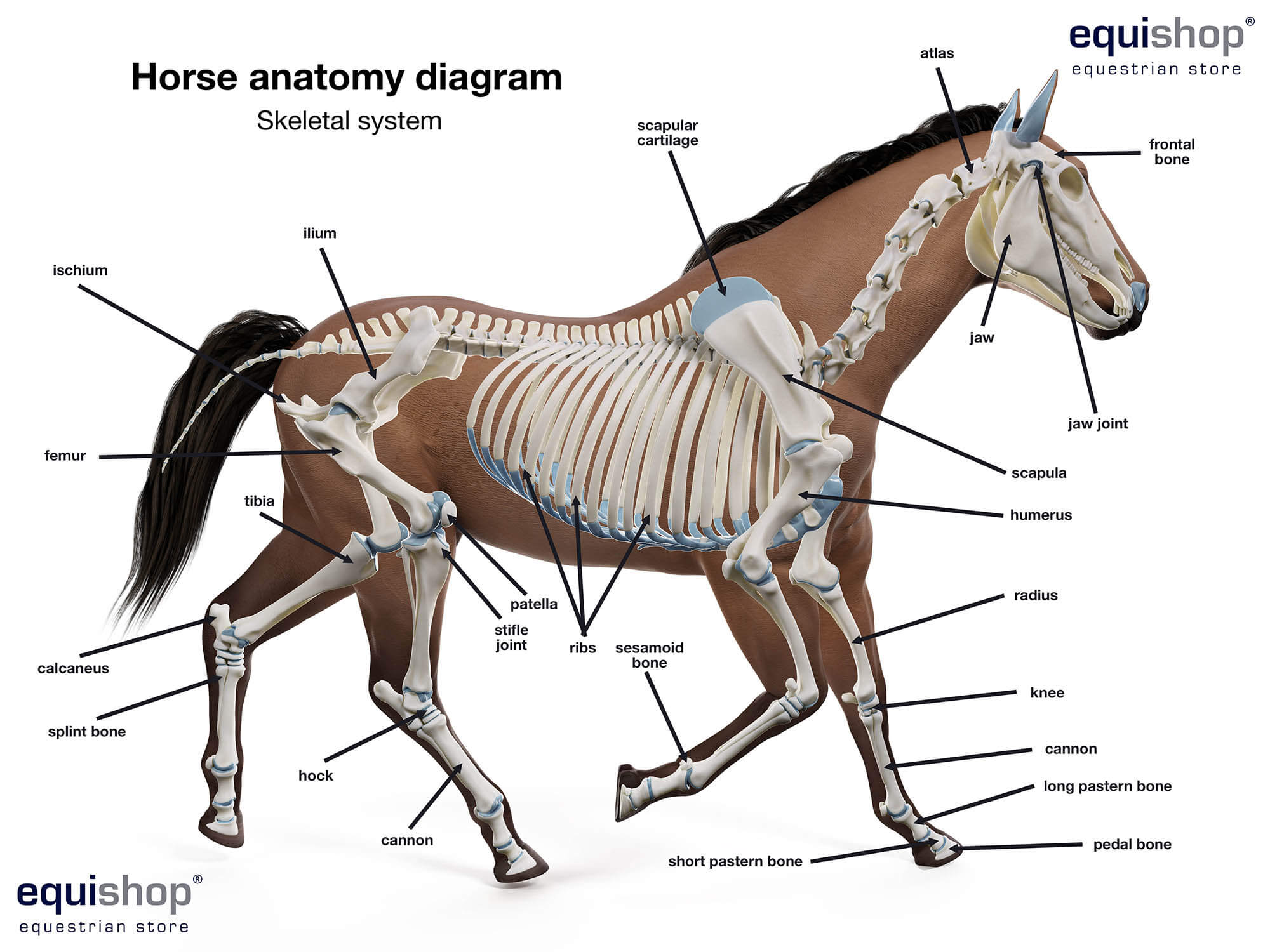
Horse Anatomy Diagrams Of Horse Body Parts Equishop Equestrian Shop
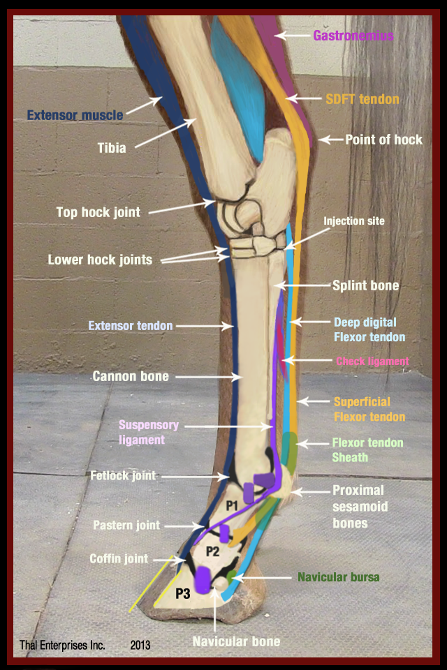
Equine Rear Leg Bones and Function · The horse leg anatomy in the rear includes the bones of the pelvis (the ilium, ischium and pubic bones), femur, tibia, ...
Horse Leg Anatomy Learn Everything You Did Not Know Medrego
#3. Horse leg anatomy (bone and muscle anatomy) Conclusion This is the summary of horse anatomy where I tried to provide valuable information. If you think you got a basic idea of the different organs system of a horse, then share this article with your friends who are also interested to learn horse anatomy.
Horse Hoof Anatomy Inside And Out
Bones In Leg Diagram / Blue Roan Pony Leg Bone Anatomy Horse Anatomy Dog Anatomy Horses. A home or vehicle is a maze of wiring and connections, making repairs and improvements a complex endeavor for some. Your legs are two of your most important body parts.

Horse Front Leg Anatomy R Oneyplays
Dog leg anatomy. First, you might have a basic idea of the different bones of the forelimb and hindlimb of a dog. Now I will provide you the few information on the other bones of dog leg anatomy with their unique features. The front leg of a dog consists of the clavicle, scapula (arm), radius and ulna (forearm), carpals, metacarpals, and phalanges (forepaw).

Horse Anatomy Mobility Health
Vitals & Anatomy - Horse Side Vet Guide from horsesidevetguide.com The majority of muscles in the leg are consi. The rotatores muscle, also referred to as the rotatores spinae, is actually a cluster of 22 small muscles in the thoracic region.
2
The tibia and the fibula make up the bones of the lower leg. The tibia is a large bone that supports the weight of the body. Its lower end forms the entire ankle bone in the horse and ox, but only the inner ankle bone in dogs, cats, pigs, and primates, where the fibula reaches the ankle on the outside.

This Is A Detailed Diagram Of A Horse S Hoof This Will Help Me Apply This To My Integument Presentation Horse Anatomy Horse Facts Horse Care
Leg Bones Diagram. The femur, or thighbone, is the longest and largest bone in the human body. Click now to learn more about the bones, muscles, and soft tissues Tibia: the largest and most medial leg bone, forming both the knee and ankle joints. The foot bones shown in this diagram are the talus, navicular, cuneiform, cuboid, metatarsals and ...
3
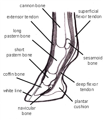
The limbs of the horse are structures made of dozens of bones, joints, muscles, tendons, and ligaments that support the weight of the equine body.
1
Horse Body Parts · Equine Forelimb Anatomy · Horse Hoof Anatomy · Horse Skeleton · Horse Teeth Anatomy · Great Anatomy Reference Books ...

Horse Leg Bones Anatomy Collection Horse Leg Anatomy Pictures Klarosa
What do you know about horses? Would you be able to pass this quiz on general equine anatomy? As it pertains to this quiz, you will be required to know the body part of the horse on the top line where the neck ends, and the back begins, what the middle joint of the back leg is called, and what is the highest point of the rump. You indeed have to take this sensational quiz.

Equine Anatomy Chart Horse Anatomy Wall Chart Amazon Com Industrial Scientific

Horse Leg Muscles And Skeleton Structure Diagram

Look Into Horse Hoof Anatomy See The Layers Of The Hoof
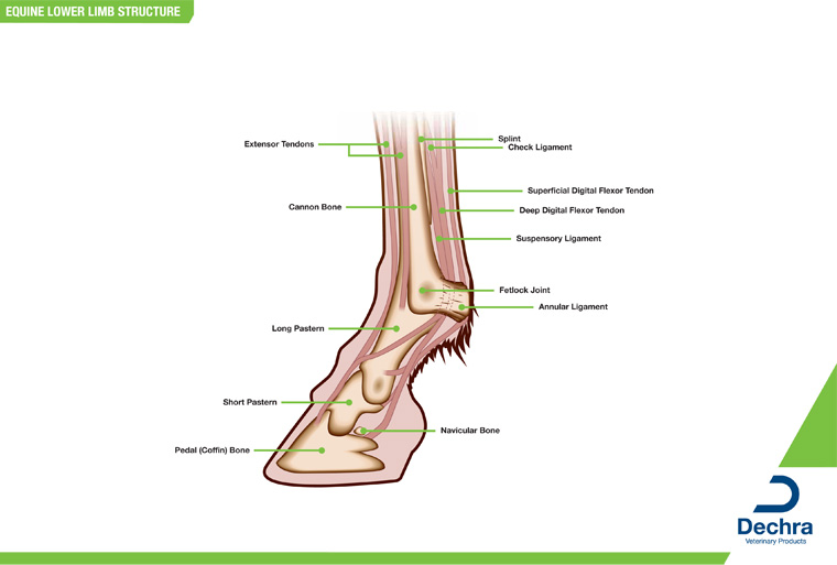
Downloads Anatomy Charts Dechra Veterinary Products
Are A Horse S Legs Really Just Fingers Below The Knee Quora
Hoof Anatomy Postcards

Horse Anatomy Diagrams Directional Terms Skeleton And Superficial Muscles

Internal Breakover Horse Anatomy Horse Care Anatomy
Equine Limb Anatomy Anatomy Drawing Diagram
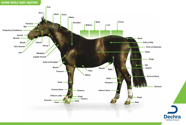
Downloads Anatomy Charts Dechra Veterinary Products

Comparing Horse And Rider Anatomy Horseclass

Equine Anatomy 3 Poster Collection Horse Organs Bone Muscles Chart
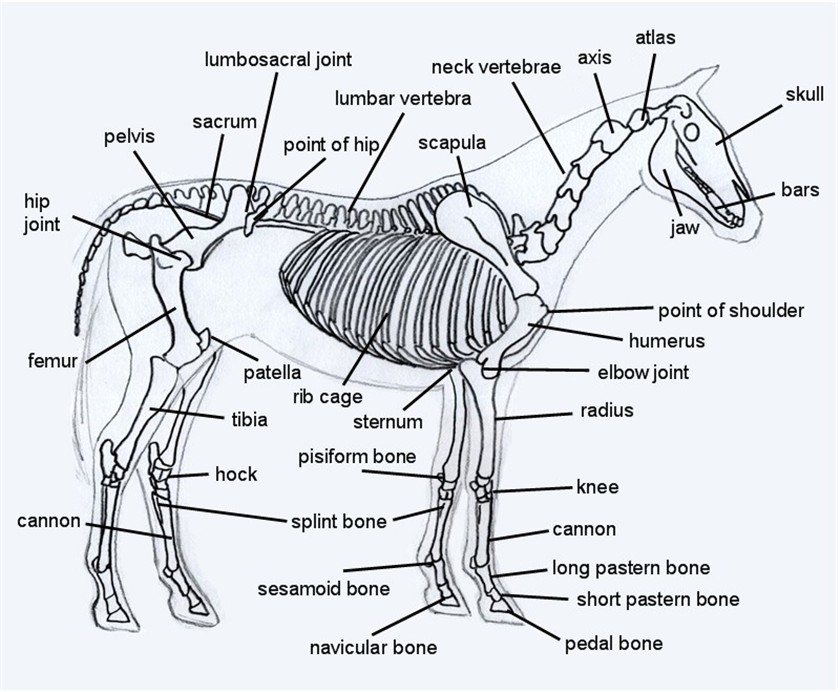
Horse Skeleton Diagram

Forever Horses Anatomy Of The Equine Hindleg By Myohodane Horse Anatomy Horse Health Large Animal Vet

Schematic Illustration Of The Musculoskeletal System Of The Horse Hind Download Scientific Diagram
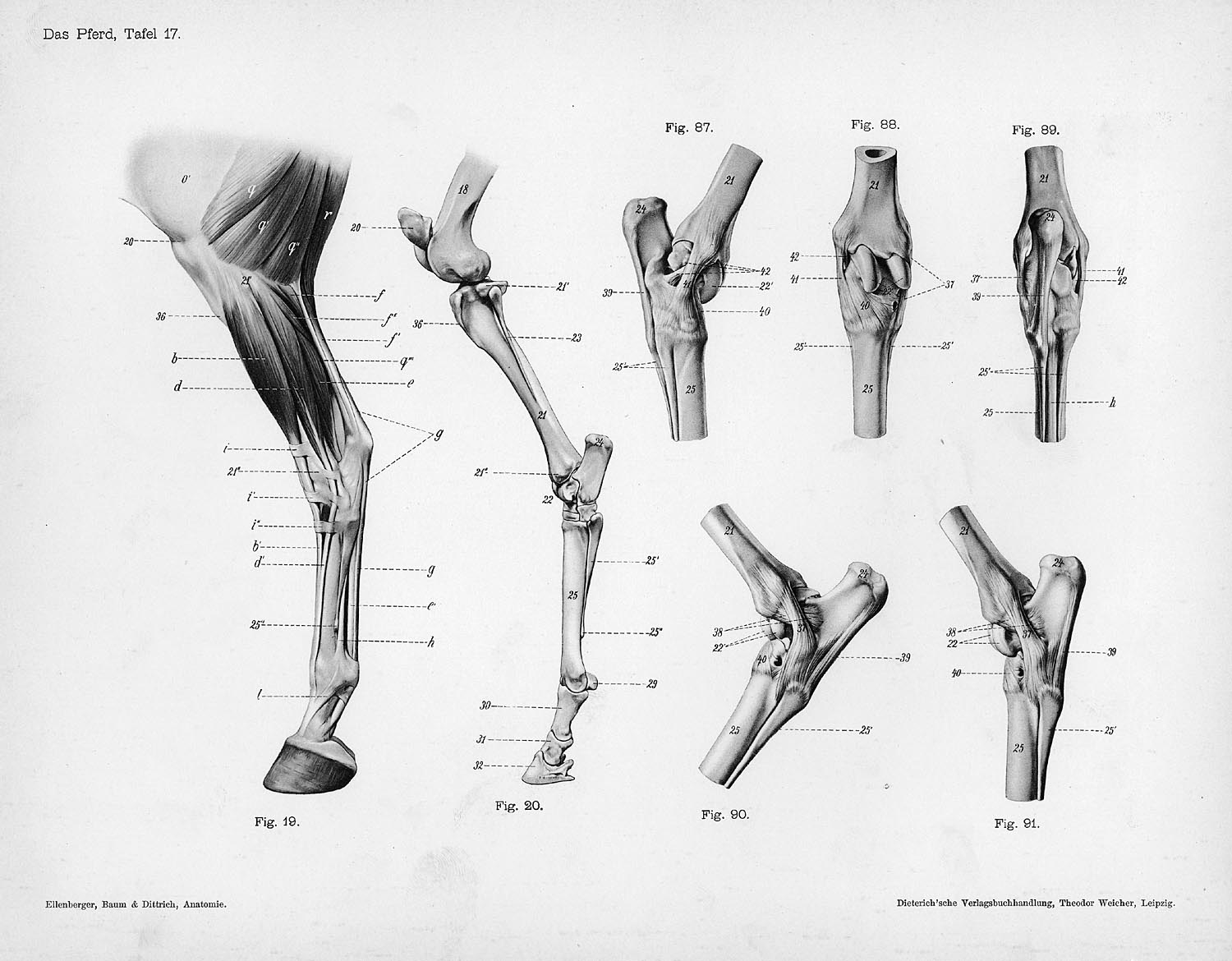
Horse Anatomy Leg Anatomy Drawing Diagram
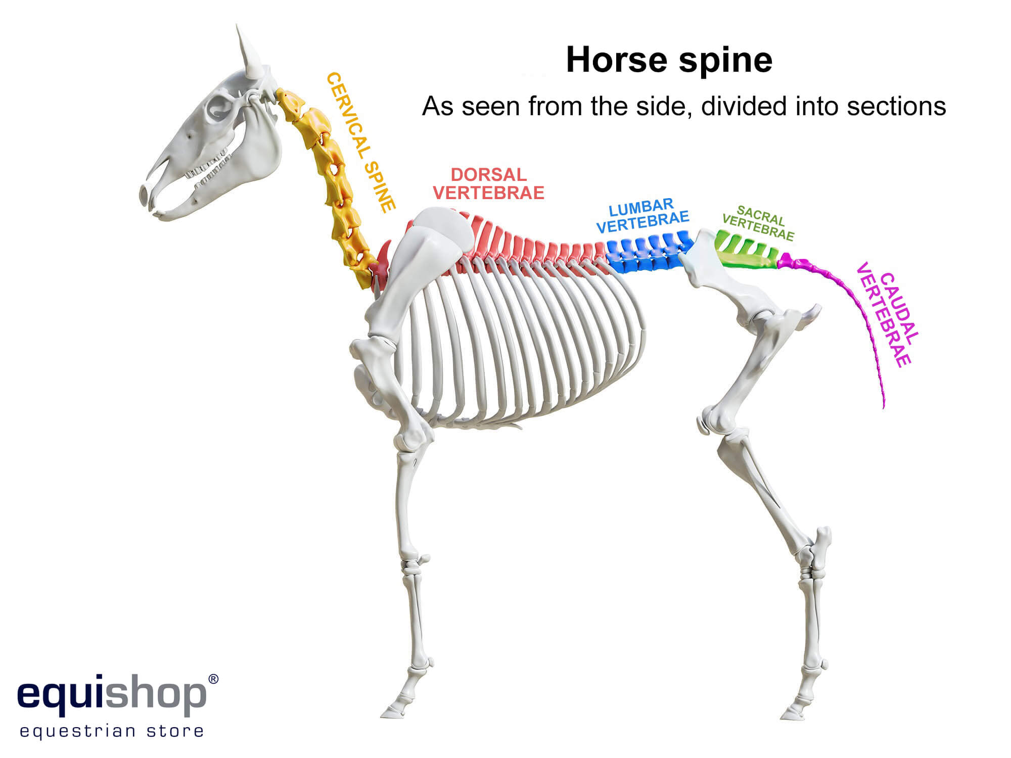
Horse Anatomy Diagrams Of Horse Body Parts Equishop Equestrian Shop
Horse Anatomy Diagrams Of Horse Body Parts Equishop Equestrian Shop
Basic Horse Anatomy For Equine Owners

How A Horse S Foot Works Aqha
0 Response to "42 horse leg anatomy diagram"
Post a Comment