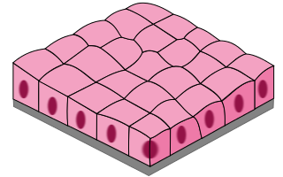
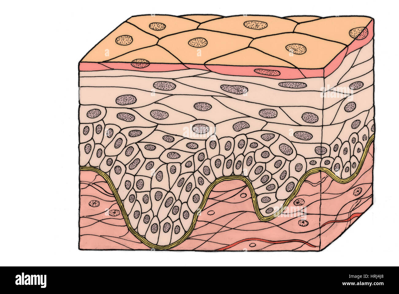
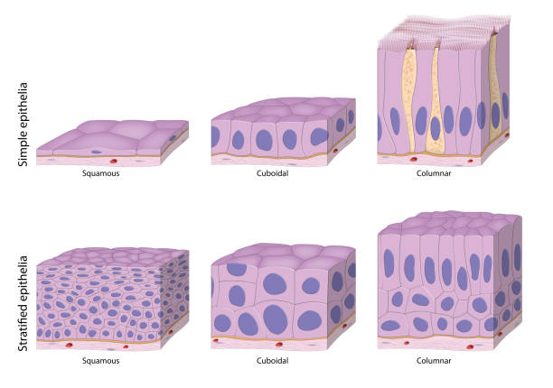
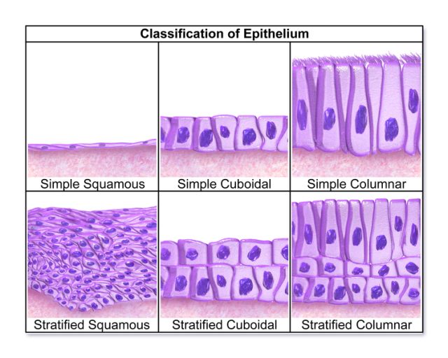
41 simple squamous epithelium diagram
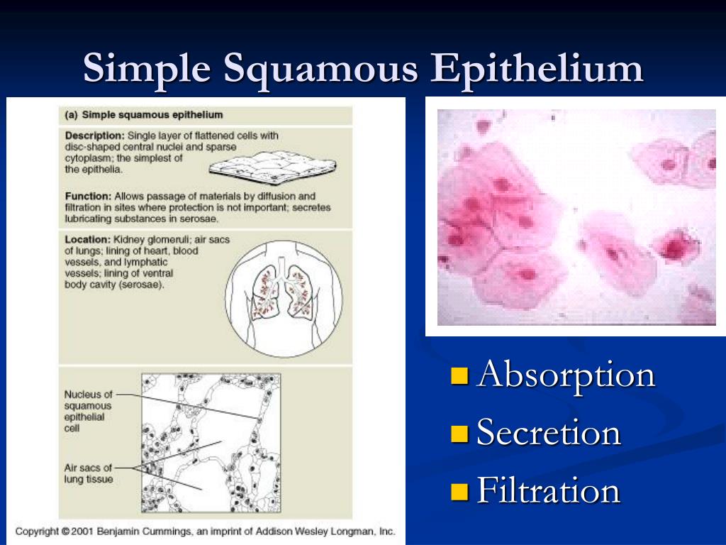
Simple squamous epithelium, isolated (400x) Buccal mucosal In the center of this image are two simple squamous epithelial cells that are still attached to each other. Notice that the location of the nucleus (nuc) is in the center of the cell. It is surrounded by the much paler cytoplasm (cyt). Squamous epithelial cells. The form and appearance of isolated squamous cells can be observed in desquamated cells from the superficial layer of the lining of the mouth (Figs. C6a and C6b).In the preparation shown here, little detail is obtained using an ordinary bright field microscope (Fig. C6a); the use of differential interference contrast (DIC) (Fig. C6b) enhances the image.
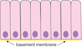
Epithelium is a tissue that lines the internal surface of the body, as well as the internal organs. Simple epithelium is one of the types of epithelium that is divided into simple columnar epithelium, simple squamous epithelium, and simple cuboidal epithelium. Bodytomy provides a labeled diagram to help you understand the structure and function of simple columnar epithelium.

Simple squamous epithelium diagram
This diagram shows that the simple squamous epithelium of the tunica adventitia layer of the heart (mesothelium) is also the visceral layer of the serous pericardium. The pericardium is a two-layered connective tissue sac that encloses the heart. The fibrous pericardium is the outer layer, and the serous pericardium is the inner layer.The space between the two layers is the pericardial cavity ... 09.11.2021 · False, because the correct statement is: In terms of structure, it is only the simple columnar, cuboidal, and squamous epithelium that is made up of a single layer of cells. Simple Squamous Epithelium (Diagram) Stratified Squamous Epithelium (Diagram) Simple Cuboidal Epithelium (Diagram) Simple Columnar Epithelium (Diagram) Ciliated Pseudostratified Columnar Epithelium (Diagram) Features. Quizlet Live. Quizlet Learn. Diagrams. Flashcards. Mobile. Help. Sign up. Help Center. Honor Code. Community Guidelines. Students.
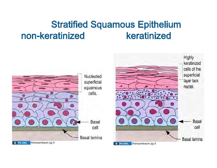
Simple squamous epithelium diagram. Stratified squamous epithelia consist of squamous as well as other cell types. They can be keratinized or non-keratinized. Though these tissues are present in the oropharynx, they are not found in alveoli, which are made of simple squamous epithelia. The thickness of this tissue in the female reproductive tract depends on hormone levels. Now look at a sagittal section of the palate (slide 115) and compare respiratory epithelium of the nasal passage View Image to the stratified squamous epithelium of the oral cavity. B. Larynx (W pg 237, 12.5) Slide 125-1 (larynx, sag. sect., H&E) View Virtual Slide. Slide 125-2 (larynx, sag. sect., H&E) View Virtual Slide 28.09.2020 · Keratinized stratified squamous epithelium is a type of stratified squamous epithelium in which the cells have a tough layer of keratin in the apical segment of cells and several layers deep to it. Keratin is a tough, fibrous intracellular protein that helps protect skin and underlying tissues from heat, microbes, and chemicals. (iii) Its mucous membrane lining is made of stratified squamous epithelial cells instead of columnar cells. It has some goblet cells which produce mucus making the food material slippery. Mucous epithelium is invaginated into submucosa forming tubular branched glands secreting an enzyme pepsin for the digestion of proteins.
A. Simple columnar epithelium. Slide 29 (small intestine) View Virtual Slide Slide 176 40x (colon, H&E) View Virtual Slide Remember that epithelia line or cover surfaces. In slide 29 and slide 176, this type of epithelium lines the luminal (mucosal) surface of the small and large intestines, respectively. Refer to the diagram at the end of this chapter for the tissue orientation and consult ... Simple Epithelium- it is composed of one layer of a cell and mostly has a secretory or an absorptive function. Compound (Stratified) Epithelium- it is made up of two or more than two layers of cells and mostly has a protective function. The glandular epithelium is made up of cuboidal or columnar cells. They are specialised for secretion. Unicellular- isolated glandular cells, e.g. goblet cells ... Apocrine (/ ˈ æ p ə k r ɪ n /) is a term used to classify exocrine glands in the study of histology.Cells which are classified as apocrine bud their secretions off through the plasma membrane producing extracellular membrane-bound vesicles. The simple squamous epithelium shown here is the outer wall of the glomerular capsule. More information about glomerular capsules and related structures is available in the section on the kidney. n = nucleus of a simple squamous epithelial cell. c = cytoplasm of a simple squamous epithelial cell.
The simple squamous epithelium location specifically exists in the lining of the blood vessels like the arteries, veins, and capillaries. It is also found lining the alveoli or air sacs within the ... Epithelium: Types of simple epithelium. Squamous. stratified squamous diagram photo of endothelial cells. Squamous means scale-like. simple squamous. Bodytomy provides a labeled diagram to help you understand the structure and Simple Columnar Epithelium: Labeled Diagram and Function. Epithelium is a tissue that lines the internal surface of the ... May 2, 2015 - simple squamous epithelium diagram - Google Search Microscope Simple Squamous Epithelium Labeled Diagram Written By MacPride Friday, December 25, 2020 Add Comment Edit. Epithelium Web Lab. What Are The Differences Of Simple And Stratified Tissue Sciencing. Epithelial Tissue Anatomy Physiology. Https Www Augusta Edu Scimath Biology Docs Animaltissues Pdf.
Epithelium (/ ˌ ɛ p ɪ ˈ θ iː l i ə m /) is one of the four basic types of animal tissue, along with connective tissue, muscle tissue and nervous tissue.It is a thin, continuous, protective layer of compactly packed cells with little intercellular matrix.Epithelial tissues line the outer surfaces of organs and blood vessels throughout the body, as well as the inner surfaces of cavities ...
Simple epithelium can be divided into 4 major classes, depending on the shapes of constituent cells. The cells found in this epithelium type are flat and thin, making simple squamous epithelium ideal for lining areas where passive diffusion of gases occur.Areas where it can be found include: skin, capillary walls, glomeruli, pericardial lining, pleural lining, peritoneal cavity lining, and ...
These labelled diagrams should closely follow the current Science courses in histology, anatomy and ... (simple squamous epithelium) ORIGIN: mesoderm lumen GENERALISED SECTION epithelium OF THE BODY connective tissue beneath epithelium connective tissue, muscle, glands, etc dermis

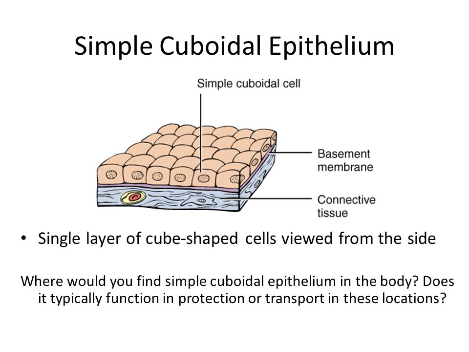
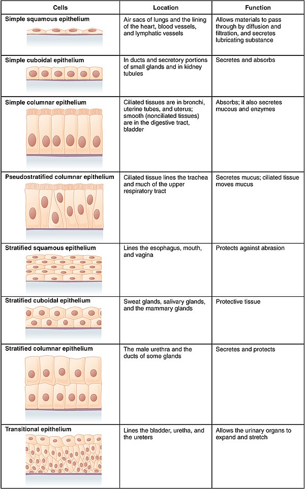
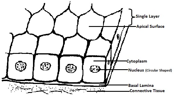
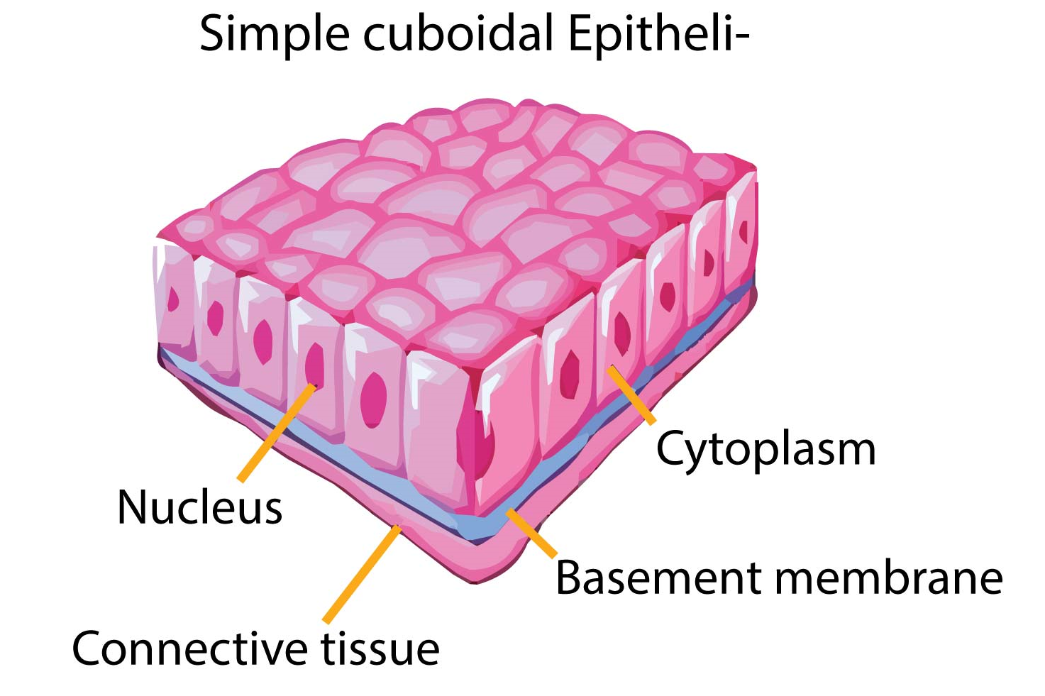
Diagram To Show The Various Kinds Of Epithelium Simple Squamous Stratified Squamous Cuboidal Columnar And Transitional Royalty Free Cliparts Vectors And Stock Illustration Image 14742327
The area beneath the stratified squamous epithelium shown in slide 33 is the dermis, which is composed of dense irregular connective tissue. In this section, the fibers clearly predominate. In this section, the fibers clearly predominate.
simple squamous epithelium. Single row of elongated cells, but some cells don't reach the free surface. pseudostratified columnar epithelium. forms walls of capillaries and air sacs of lungs. simple squamous epithelium. Provides lining of urethra of males and parts of pharynx. stratified columnar epithelium . provides abrasion protection of skin epidermis and oral cavity. stratified squamous ...
27.09.2020 · Simple squamous epithelium is a type of simple epithelium that is formed by a single layer of cells on a basement membrane. It is a type of epithelium formed by a single layer of squamous or flat cells present on a thin extracellular layer, called the basement membrane. This epithelium is also termed the pavement epithelium because the cells appear like tiles on a floor when viewed from the ...
Simple squamous epithelia are found both in alveoli and capillaries and their flat, thin, single-layered structure is important for the exchange of gases between the lungs and blood. Though simple squamous epithelia are involved in the filtration of nitrogenous waste products, the process occurs in the kidney, not the brain.
Simple squamous epithelium, c.s. (40X) Kidney cortex . This very tall image shows almost the entire thickness of the kidney. Your goal is to find and learn to recognize simple squamous epithelium on a slide similar to this. The easiest place to find this tissue is the glomerular capsule (don't worry, you don't have to know what that is to find ...
Simple Epithelia Simple Squamous Epithelium (Figure 4.3a) A simple squamous epithelium is a single layer of flat cells. When viewed from above, the closely fitting cells resemble a tiled floor. When viewed in lateral section, they resemble fried eggs seen from the side. Thin and often permeable, this type
Histology diagram of simple squamous epithelium histology diagram. Both surface and side view has been demonstrated in this video. A simple squamous epitheli...
Browse 56 simple squamous epithelium stock illustrations and vector graphics available royalty-free, or search for simple columnar epithelium or stratified squamous epithelium to find more great stock images and vector art. Capillary. blood vessel. labelled Capillary. blood vessel. labelled.
12.08.1996 · Mesothelium = the simple squamous epithelium lining body cavities and mesenteries. Slide 2 High power view of endothelial cells lining a small blood vessel cut in cross-section. (You see just the nuclei - the cytoplasm between them is extremely flat.) Endothelium = the simple squamous epithelium lining blood vessels. Slide 3 Low power view of larger vessels, showing endothelial nuclei …
Sep 07, 2021 · Ciliated epithelium is an important tissue found in various parts of the body and aids in everyday health. Explore what ciliated epithelial is and its function, its structure using a diagram, why ...
23.08.2021 · Unlike simple epithelium, just the outermost layer of the tissue is exposed to the lumen at the apical surface of stratified epithelium. Cell junctions and adhesions connect all of the other sides of the cells to each other. These cells have multiple desmosomes and other adhesins, and as they approach closer to the surface, their shape becomes more uneven, then flattens. They become dehydrated ...
Merocrine (or eccrine) is a term used to classify exocrine glands and their secretions in the study of histology.A cell is classified as merocrine if the secretions of that cell are excreted via exocytosis from secretory cells into an epithelial-walled duct or ducts and then onto a bodily surface or into the lumen.. Merocrine is the most common manner of secretion.
The parietal layer of Bowman's capsule is also a simple squamous epithelium which transitions to cuboidal epithelium of the proximal convoluted tubule at the urinary pole #210 . Look around under low power to find glomeruli sectioned through the vascular pole. Near the vascular pole will be the distal tubule of the same nephron.
A simple squamous epithelium, also known as pavement epithelium, and tessellated epithelium is a single layer of flattened, polygonal cells in contact with the basal lamina (one of the two layers of the basement membrane) of the epithelium. This type of epithelium is often permeable and occurs where small molecules need to pass quickly through membranes via filtration or diffusion.
What is the structure of the simple squamous? Tightly packed cells. What is the shape of these cells? Flat and thin. Where are these cells located? Lymph vessels, capillaries, and alveoli (tiny air sacs in the lungs). What are the functions of these cells? (Absorption, secretion, and filtration). Forms the walls of the capillaries, lines the ...
Simple squamous epithelium is the tissue that creates from one layer of squamous cells which line surfaces. The squamous cells are thin, large, and flat, and consisting of around nucleus. These tissues have polarity like other epithelial cells and consist of a distinct apical surface with special membrane proteins.
Simple squamous epithelia are a single layer of flattened cells that because of their thinness, facilitate exchange of gases and are found in the lung and blood vessels. Simple cuboidal and columnar are taller and are actively involved in absorption and secretion. Both of these activities require more organelles for protein secretion (ER, Golgi) and/or energy generation (mitochondria), making ...

13 Stratified Squamous Epithelium Ideas Stratified Squamous Epithelium Squamous Anatomy And Physiology
Simple Squamous Epithelium (Diagram) Stratified Squamous Epithelium (Diagram) Simple Cuboidal Epithelium (Diagram) Simple Columnar Epithelium (Diagram) Ciliated Pseudostratified Columnar Epithelium (Diagram) Features. Quizlet Live. Quizlet Learn. Diagrams. Flashcards. Mobile. Help. Sign up. Help Center. Honor Code. Community Guidelines. Students.
09.11.2021 · False, because the correct statement is: In terms of structure, it is only the simple columnar, cuboidal, and squamous epithelium that is made up of a single layer of cells.
This diagram shows that the simple squamous epithelium of the tunica adventitia layer of the heart (mesothelium) is also the visceral layer of the serous pericardium. The pericardium is a two-layered connective tissue sac that encloses the heart. The fibrous pericardium is the outer layer, and the serous pericardium is the inner layer.The space between the two layers is the pericardial cavity ...

What Are The Various Forms Of Cells Of Epithelial Tissue Describe Briefly From Science Tissues Class 9 Cbse

Pseudostratified Columnar Epithelium Simple Columnar Epithelium Trachea Organ Others Purple Angle Violet Png Pngwing













0 Response to "41 simple squamous epithelium diagram"
Post a Comment