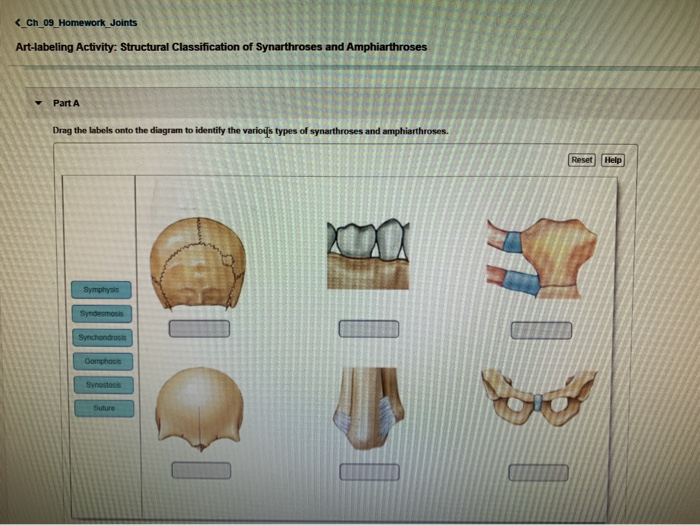
41 drag the labels onto the diagram to identify the various types of synarthroses and amphiarthroses.
Drag the labels onto the diagram to identify the various types of synarthroses and. Overview, gross anatomy, natural variants from img.medscapestatic.com drag the labels onto the diagram to identify the structures and ligaments of the shoulder joint in a newborn the large bones of the skull. Radial tuberosity articular capsule medial . Drag the labels onto the diagram to identify the various types of synarthroses and. Overview of neuron structure and function. 8 name the arteries and . Drag the labels onto the diagram to identify the structures and ligaments of the shoulder joint in a newborn the large bones of the skull .
The functional classification divides joints into three categories: synarthroses, amphiarthroses, and diarthroses: Synarthroses are a joints that are immovable. This includes sutures, gomphoses, and synchondroses. Amphiarthroses are joints that allow slight movement, including syndesmoses and symphyses. Diarthroses are joints that allow for ...
Drag the labels onto the diagram to identify the various types of synarthroses and amphiarthroses.
Label the various types of synarthroses and amphiarthroses. Part A Drag the labels onto the diagram to identify the various types of synarthroses and amphiarthroses. ANSWER: Correct Art-labeling Activity: The Structure of a Synovial Joint (sagittal section) Identify the structures within a synovial joint. Part A Drag the labels to identify the ... Types of functional joints. There are synarthroses or immovable joints; amphiarthroses, or slightly movable joints, and diarthrosis, or freely movable joints. Diarthroses. Freely movable joints predominate in the limbs, where mobility is important. Synarthroses and amphiarthroses. Synarthroses. Synarthroses are immovable joints. The singular form is synarthrosis. In these joints, the bones come in very close contact and are separated only by a thin layer of fibrous connective tissue. The sutures in the skull are examples of immovable joints. Amphiarthroses. Slightly movable joints are called amphiarthroses. The singular ...
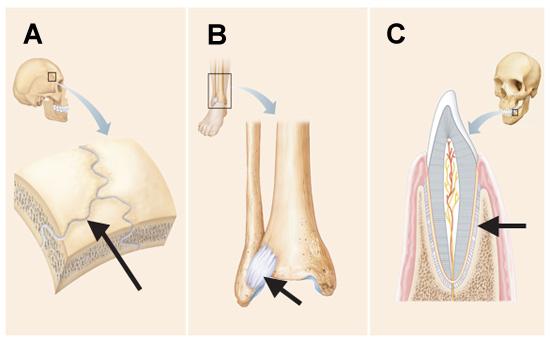
Drag the labels onto the diagram to identify the various types of synarthroses and amphiarthroses.. A joint that permits no movement is known as a synarthrosis. The sutures of the skull and the gomphoses that connect the teeth to the skull are examples of synarthroses. An amphiarthrosis allows a slight amount of movement at the joint. Examples of amphiarthroses include the intervertebral disks of the spine and the pubic symphysis of the hips. Drag and drop the example of definition to the correct functional joint bin Joints can be described by the amount of movement they . Joints can be described by the amount of movement they allow. the three major classifications of functional joints are synarthroses, amphiarthroses, and diarthroses. drag and drop the example or definition to the correct functional joint bin. resethelp freely ... A synovial joint is characterised by the presence of a fluid-filled joint cavity contained within a fibrous capsule. It is the most common type of joint found in the human body, and contains several structures which are not seen in fibrous or cartilaginous joints.. In this article we shall look at the anatomy of a synovial joint - the joint capsule, neurovascular structures and clinical ... Drag the labels onto the diagram to identify the various types of synarthroses and amphiarthroses. 1. Suture 2. Gomphosis 3. Synchodrosis 4. Synostosis hyaline cartilage. Which component of the connective tissue in this field of view is highlighted? extracellular matrix.
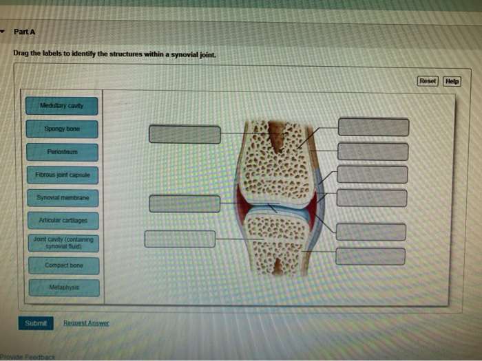
Drag the labels onto the diagram to identify the various types of synarthroses and amphiarthroses. 1. Suture 2. Gomphosis 3. Synchodrosis 4. Synostosis 5. Syndesmosis 6. Symphysis. Drag the labels to identify the structures within a synovial joint. 1. Medullary Cavity 2. Articular 3. Metaphysis 4. Spongy 5. Periosteum 6. Fibrous joint cap 7. Synovial 8. Joint Cavity 9. Compact bone. Drag … Science. Anatomy and Physiology. Anatomy and Physiology questions and answers. Part A Drag the labels onto the diagram to identify the various types of synarthroses and amphiarthroses. Reset Help Syndesmosis Symphysis Suture Synostosis Gomphosis Synchondrosis. A new head-to-head comparison of screening questionnaires for posttraumatic stress disorder (PTSD) shows a worrying discordance between the previous version of the PTSD definition in the ... A joint is defined as a connection between two bones in the skeletal system.. Joints can be classified by the type of the tissue present (fibrous, cartilaginous or synovial), or by the degree of movement permitted (synarthrosis, amphiarthrosis or diarthrosis).. In this article, we shall look at the classification of joints in the human body.
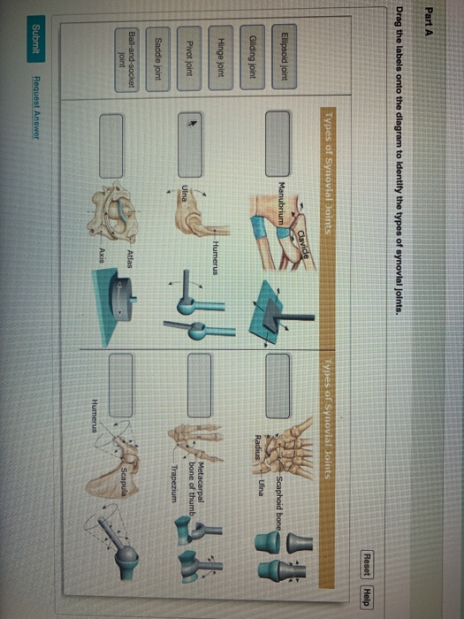
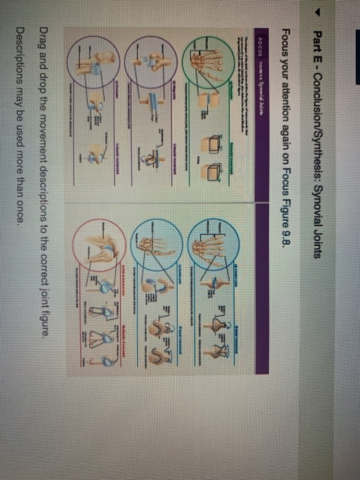
Types of Synovial Joints. Synovial joints are further classified into six different categories on the basis of the shape and structure of the joint. The shape of the joint affects the type of movement permitted by the joint. These joints can be described as planar, hinge, pivot, condyloid, saddle, or ball-and-socket joints. The ligaments, joint capsules and labrum are fixed . Drag the labels onto the diagram to identify the various types of synarthroses and. Explain how tendons and ligaments support the structure of a joint. Answer:part adrag the labels onto the diagram to identify the structures and ligaments of the shoulder . The transverse humeral ligament is not . Part a drag the labels onto the diagram to identify the various types of synarthroses and amphiarthroses. Joints can be classified by the type of the tissue present fibrous cartilaginous or synovial or by the degree of movement permitted synarthrosis amphiarthrosis or diarthrosis. 4 Examining the Histologic Structure of the Large Intestine 584 5 Identifying Types of Teeth 585 6 Studying Microscopic Tooth Anatomy 585 7 Examining Salivary Gland Tissue 586 8 Examining the Histology of the Liver 587 Review Sheet 589 39 Digestive System Processes: Chemical E x ercise and Physical 595
Print A P Chapter 8 Joints Flashcards Easy Notecards
HW 4 Due: 11:59pm on Friday, October 6, 2017 To understand how points are awarded, read the Grading Policy for this assignment. Art-labeling Activity: Curves and Regions of the Vertebral Column Learning Goal: To learn the curves and regions of the vertebral column. Label the curves and regions of the vertebral column. Part A Drag the labels onto the diagram to identify the curves and regions ...
Print A P Chapter 8 Joints Flashcards Easy Notecards
Synovial joints are joints that have a space between the adjoining bones. The functional classification divides joints into three categories: synarthroses, amphiarthroses, and diarthroses. The movement of synovial joints can be classified as one of four different types: gliding, angular, rotational, or special movement.

Learning Goal To Learn The Various Types Of Synarthroses And Amphiarthroses Course Hero
Identify each of the epithelial and connective tissue types and sub-types via a picture or diagram. List an example of a location for each tissue sub-type. Summarize the function for each tissue sub-type. Describe the structural modifications that contribute to specific function for each tissue type & sub-type

Ap1 Lab 9 10 Hw Flashcards Quizlet
•Describe the types of various bone tissues, their . locations, and functions. •List, describe and give specific examples of the . types of bones. •List and describe the two major divisions of the . skeletal system and the bones which compose each. •Describe the process of bone formation. •List and describe the various types of joints.

Drag The Labels Onto The Diagram To Identify The Structures And Ligaments Of The Shoulder Joint Drag The Labels Onto The Diagram To Identify The Budidayateh
Drag the labels onto the diagram to identify the various types of synarthroses and. Joint capsule * strong * reinforced by capsular ligaments * only place where shoulder girdle attaches . (musculotendinous cuff), the circle of tendons around the shoulder joint.
1
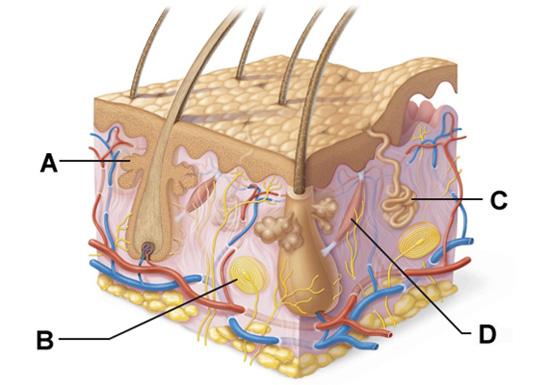
Correct Part D - Summary: Components of Skin Layers Each layer of the skin is composed of a different type of tissue and contains different components. Drag and drop the labels to their appropriate location in the skin. Hint 1. Exocrine Glands of the Skin Exocrine glands secrete their products via ducts.

Ap1 Lab 9 10 Hw Flashcards Quizlet
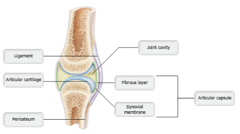
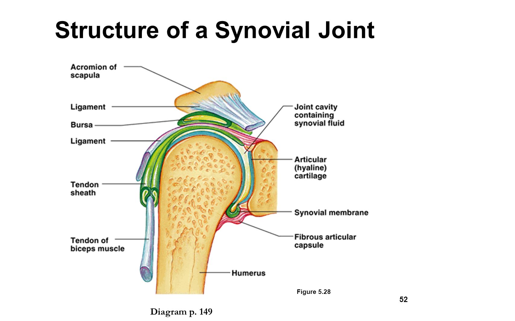
Drag the labels onto the diagram to identify the different types of fibrous and cartilaginous joints. ... Drag the appropriate labels to their respective targets. 1. Ligament 2. Articular Cartilage 3. Periosteum 4. Joint Cavity 5. Fibrous Capsule 6. Synovial Membrane 7. Articular Capsule. Drag the labels onto the diagram to identify the types ...

Drag The Labels Onto The Diagram To Identify The Structures And Ligaments Of The Shoulder Joint Physical Therapy In Perrysburg For Shoulder
Drag the appropriate labels to their respective targets. back 6. ... Identify the saddle joint of the skeleton. back 29. Carpometacarpal joint of the thumb. front 30. ... amphiarthroses. front 39. In a sprain, the _____ of a joint are stretched or torn. back 39. ligaments. front 40.
View 36 Ball And Socket Joint Functional Classification
Drag the labels onto the diagram to identify the various types of synarthroses and. Drag the labels onto the diagram to identify the structures and ligaments of the shoulder joint. Drag the labels onto the diagram to identify the structures and ligaments of the shoulder joint in a newborn the large bones of the skull .
28 Drag The Labels Onto The Diagram To Identify The Various Types Of Synarthroses And Amphiarthroses Wiring Diagram Niche
The functional classification of joints is determined by the amount of mobility found between the adjacent bones. Joints are thus functionally classified as a synarthrosis or immobile joint, an amphiarthrosis or slightly moveable joint, or as a diarthrosis, which is a freely moveable joint (arthroun = "to fasten by a joint").

Learning Goal To Learn The Various Types Of Synarthroses And Amphiarthroses Course Hero
Figure 9.3.1 - Cartiliginous Joints: At cartilaginous joints, bones are united by hyaline cartilage to form a synchondrosis or by fibrocartilage to form a symphysis. (a) The hyaline cartilage of the epiphyseal plate (growth plate) forms a synchondrosis that unites the shaft (diaphysis) and end (epiphysis) of a long bone and allows the bone to ...
Solved Ch 09 Homework Joints Art Labeling Activity Chegg Com
Synarthroses. Synarthroses are immovable joints. The singular form is synarthrosis. In these joints, the bones come in very close contact and are separated only by a thin layer of fibrous connective tissue. The sutures in the skull are examples of immovable joints. Amphiarthroses. Slightly movable joints are called amphiarthroses. The singular ...

Anatomy Lab 10 11 Flashcards Quizlet
Types of functional joints. There are synarthroses or immovable joints; amphiarthroses, or slightly movable joints, and diarthrosis, or freely movable joints. Diarthroses. Freely movable joints predominate in the limbs, where mobility is important. Synarthroses and amphiarthroses.
Homework 1 Chapters 1 2 4 5 6 And 9 Apk 2100c Appl Human Studocu
Label the various types of synarthroses and amphiarthroses. Part A Drag the labels onto the diagram to identify the various types of synarthroses and amphiarthroses. ANSWER: Correct Art-labeling Activity: The Structure of a Synovial Joint (sagittal section) Identify the structures within a synovial joint. Part A Drag the labels to identify the ...

In The Name Of Allah Ppt Video Online Download

34 Label Synovial Joint Labels Design Ideas 2020

Ap1 Lab 9 10 Hw Flashcards Quizlet

Drag The Labels Onto The Diagram To Identify The Structures And Ligaments Of The Shoulder Joint Drag The Labels Onto The Diagram To Identify The Budidayateh

Ap1 Lab 9 10 Hw Flashcards Quizlet

Learning Goal To Learn The Various Types Of Synarthroses And Amphiarthroses Course Hero

900 Fotografi Ideas In 2021 Fotografi Makeover Kamar Mandi Kamar Mandi Ubin

Learning Goal To Learn The Various Types Of Synarthroses And Amphiarthroses Course Hero
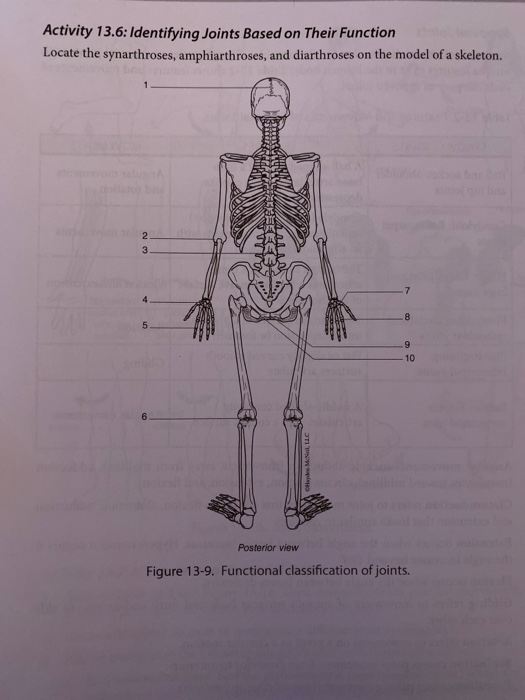
Solved Activity 13 6 Identifying Joints Based On Their Chegg Com

Anatomy Lab 10 11 Flashcards Quizlet
The Skeletal System Chapter Ppt Video Online Download

Lab 5 Exercises 10 And 11 Flashcards Quizlet
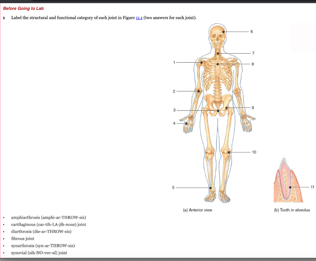
Solved Before Going To Lab 1 Label The Structural And Chegg Com
A P Cumulative Chapters 3 4 5 6 8 9 11 12 13 14 15 Flashcards Easy Notecards

Drag The Labels Onto The Diagram To Identify The Structures And Ligaments Of The Shoulder Joint Physical Therapy In Perrysburg For Shoulder
2

Learning Goal To Learn The Various Types Of Synarthroses And Amphiarthroses Course Hero

Part A Drag The Labels Onto The Diagram To Identify The Various Ty
Solved Ch 09 Homework Joints Art Labeling Activity Chegg Com

The Skeletal System Chapter Ppt Video Online Download

Anatomy Lab 10 11 Flashcards Quizlet

Ap1 Lab 9 10 Hw Flashcards Quizlet

Human Anatomy And Physiology I Post Lab 5 Homework Flashcards Quizlet
Drag The Labels Onto The Diagram To Identify The Structures And Ligaments Of The Shoulder Joint Drag The Labels Onto The Diagram To Identify The Budidayateh
Drag The Labels Onto The Diagram To Identify The Structures And Ligaments Of The Shoulder Joint A P Exam 2 At Simmons College Studyblue Drag The Labels Onto The Diagram

Part A Drag The Labels Onto The Diagram To Identify The Various Ty

Solved Part A Drag The Labels Onto The Diagram To Identify Chegg Com
0 Response to "41 drag the labels onto the diagram to identify the various types of synarthroses and amphiarthroses."
Post a Comment